 Movie
Movie Controller

[English] 日本語
 Yorodumi
- PDB-4onc: Crystal Structure of Mycobacterium Tuberculosis Decaprenyl Diphos... -
+ Open data

- Basic information
| Entry | Database: PDB / ID: 4onc | ||||||
|---|---|---|---|---|---|---|---|
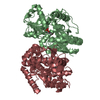
| Title | Crystal Structure of Mycobacterium Tuberculosis Decaprenyl Diphosphate Synthase in Complex with BPH-640 | ||||||
 Components Components | Decaprenyl diphosphate synthase | ||||||
 Keywords Keywords | TRANSFERASE / polyprenyl transferase / decaprenyl diphosphate synthase | ||||||
| Function / homology |  Function and homology information Function and homology informationtrans,polycis-decaprenyl diphosphate synthase / Z-farnesyl diphosphate synthase activity / ditrans,polycis-polyprenyl diphosphate synthase [(2E,6E)-farnesyl diphosphate specific] / all-trans-nonaprenyl-diphosphate synthase (geranyl-diphosphate specific) activity / ditrans,polycis-undecaprenyl-diphosphate synthase [(2E,6E)-farnesyl-diphosphate specific] activity / polyprenol biosynthetic process / prenyltransferase activity / manganese ion binding / magnesium ion binding / plasma membrane / cytosol Similarity search - Function | ||||||
| Biological species |   Mycobacterium tuberculosis (bacteria) Mycobacterium tuberculosis (bacteria) | ||||||
| Method |  X-RAY DIFFRACTION / SYNCHROTRON / MOLECULAR REPLACEMENT / Resolution: 1.83 Å X-RAY DIFFRACTION / SYNCHROTRON / MOLECULAR REPLACEMENT / Resolution: 1.83 Å | ||||||
 Authors Authors | Feng, X. / Chan, H.C. / Ko, T.P. | ||||||
 Citation Citation | Journal: J.Am.Chem.Soc. / Year: 2014 Title: Structure and Inhibition of Tuberculosinol Synthase and Decaprenyl Diphosphate Synthase from Mycobacterium tuberculosis Authors: Chan, H.C. / Feng, X. / Ko, T.P. / Huang, C.H. / Hu, Y. / Zheng, Y. / Bogue, S. / Nakano, C. / Hoshino, T. / Zhang, L. / Lv, P. / Liu, W. / Crick, D.C. / Liang, P.H. / Wang, A.H. / Oldfield, E. / Guo, R.T. | ||||||
| History |
|
- Structure visualization
| Structure viewer | Molecule: MolmilJmol/JSmol |
|---|
- Downloads & links
-Download
| PDBx/mmCIF format | 4onc.cif.gz | 134.6 KB | Display | PDBx/mmCIF format |
|---|---|---|---|---|
| PDB format | pdb4onc.ent.gz | 104.2 KB | Display | PDB format |
| PDBx/mmJSON format | 4onc.json.gz | Tree view | PDBx/mmJSON format | |
| Others |  Other downloads Other downloads |
-Validation report
| Arichive directory | https://data.pdbj.org/pub/pdb/validation_reports/on/4oncftp://data.pdbj.org/pub/pdb/validation_reports/on/4onc | HTTPS FTP |
|---|
-Related structure data
| Related structure data |  3wqkC  3wqlC  3wqmC  3wqnC  4kt8C  2vg4S S: Starting model for refinement C: citing same article ( |
|---|---|
| Similar structure data |


-Links
 PDBj
PDBj- Assembly
| Deposited unit | 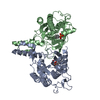
| ||||||||
|---|---|---|---|---|---|---|---|---|---|
| 1 |
| ||||||||
| Unit cell |
|
-Components
| #1: Protein | Mass: 32460.809 Da / Num. of mol.: 2 / Fragment: UNP RESIDUES 13-296 Source method: isolated from a genetically manipulated source Source: (gene. exp.) Mycobacterium tuberculosis (bacteria) / Gene: Rv2361c / Production host: References: UniProt: P60479, UniProt: P9WFF7*PLUS, trans,polycis-decaprenyl diphosphate synthase, ditrans,polycis-polyprenyl diphosphate synthase [(2E,6E)-farnesyl diphosphate specific] #2: Chemical |   Mass: 420.290 Da / Num. of mol.: 2 / Source method: obtained synthetically / Formula: C19H18O7P2 Mass: 420.290 Da / Num. of mol.: 2 / Source method: obtained synthetically / Formula: C19H18O7P2#3: Water | ChemComp-HOH / |  Mass: 18.015 Da / Num. of mol.: 437 / Source method: isolated from a natural source / Formula: H2O Mass: 18.015 Da / Num. of mol.: 437 / Source method: isolated from a natural source / Formula: H2O |
|---|
-Experimental details
-Experiment
| Experiment | Method: X-RAY DIFFRACTION / Number of used crystals: 1 |
|---|
- Sample preparation
| Crystal | Density Matthews: 2.51 Å3/Da / Density % sol: 50.96 % |
|---|---|
| Crystal grow | Temperature: 298 K / Method: vapor diffusion, sitting drop / pH: 7.5 Details: 0.1M HEPES, 10% GLYCEROL, 25% PEG 400, 7% PEG 3000, pH 7.5, VAPOR DIFFUSION, SITTING DROP, temperature 298K |
-Data collection
| Diffraction | Mean temperature: 298 K |
|---|---|
| Diffraction source | Source: SYNCHROTRON / Site: APS  / Beamline: 21-ID-G / Wavelength: 0.97857 / Beamline: 21-ID-G / Wavelength: 0.97857 |
| Detector | Type: MARMOSAIC 300 mm CCD / Detector: CCD / Date: Jun 1, 2012 |
| Radiation | Monochromator: C(111) / Protocol: SINGLE WAVELENGTH / Monochromatic (M) / Laue (L): M / Scattering type: x-ray |
| Radiation wavelength | Wavelength: 0.97857 Å / Relative weight: 1 |
| Reflection | Resolution: 1.83→50 Å / Num. obs: 58370 / % possible obs: 100 % / Observed criterion σ(I): 0 |
| Reflection shell | Resolution: 1.83→1.86 Å / % possible all: 100 |
- Processing
| Software |
| ||||||||||||||||||||||||||||||||||||||||||||||||||||||||||||||||||||||||||||||||||||||||||||||||||||||||||||||||||||||||||||||||||||||||||||||||||||||||||||||||||||||||||||||||||||||
|---|---|---|---|---|---|---|---|---|---|---|---|---|---|---|---|---|---|---|---|---|---|---|---|---|---|---|---|---|---|---|---|---|---|---|---|---|---|---|---|---|---|---|---|---|---|---|---|---|---|---|---|---|---|---|---|---|---|---|---|---|---|---|---|---|---|---|---|---|---|---|---|---|---|---|---|---|---|---|---|---|---|---|---|---|---|---|---|---|---|---|---|---|---|---|---|---|---|---|---|---|---|---|---|---|---|---|---|---|---|---|---|---|---|---|---|---|---|---|---|---|---|---|---|---|---|---|---|---|---|---|---|---|---|---|---|---|---|---|---|---|---|---|---|---|---|---|---|---|---|---|---|---|---|---|---|---|---|---|---|---|---|---|---|---|---|---|---|---|---|---|---|---|---|---|---|---|---|---|---|---|---|---|---|
| Refinement | Method to determine structure: MOLECULAR REPLACEMENT Starting model: 2VG4 Resolution: 1.83→49.73 Å / Cor.coef. Fo:Fc: 0.97 / Cor.coef. Fo:Fc free: 0.958 / SU B: 2.36 / SU ML: 0.072 / Cross valid method: THROUGHOUT / ESU R: 0.11 / ESU R Free: 0.111 / Stereochemistry target values: MAXIMUM LIKELIHOOD / Details: HYDROGENS HAVE BEEN USED IF PRESENT IN THE INPUT
| ||||||||||||||||||||||||||||||||||||||||||||||||||||||||||||||||||||||||||||||||||||||||||||||||||||||||||||||||||||||||||||||||||||||||||||||||||||||||||||||||||||||||||||||||||||||
| Solvent computation | Ion probe radii: 0.8 Å / Shrinkage radii: 0.8 Å / VDW probe radii: 1.2 Å / Solvent model: MASK | ||||||||||||||||||||||||||||||||||||||||||||||||||||||||||||||||||||||||||||||||||||||||||||||||||||||||||||||||||||||||||||||||||||||||||||||||||||||||||||||||||||||||||||||||||||||
| Displacement parameters | Biso mean: 28.08 Å2
| ||||||||||||||||||||||||||||||||||||||||||||||||||||||||||||||||||||||||||||||||||||||||||||||||||||||||||||||||||||||||||||||||||||||||||||||||||||||||||||||||||||||||||||||||||||||
| Refinement step | Cycle: LAST / Resolution: 1.83→49.73 Å
| ||||||||||||||||||||||||||||||||||||||||||||||||||||||||||||||||||||||||||||||||||||||||||||||||||||||||||||||||||||||||||||||||||||||||||||||||||||||||||||||||||||||||||||||||||||||
| Refine LS restraints |
|