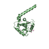
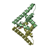
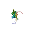
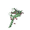
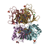
A: Protein of unknown function with a cupin-like fold B: Protein of unknown function with a cupin-like fold C: Protein of unknown function with a cupin-like fold D: Protein of unknown function with a cupin-like fold E: Protein of unknown function with a cupin-like fold F: Protein of unknown function with a cupin-like fold G: Protein of unknown function with a cupin-like fold H: Protein of unknown function with a cupin-like fold hetero molecules
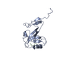
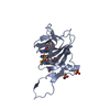
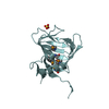
A: Protein of unknown function with a cupin-like fold B: Protein of unknown function with a cupin-like fold C: Protein of unknown function with a cupin-like fold D: Protein of unknown function with a cupin-like fold hetero molecules
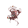
E: Protein of unknown function with a cupin-like fold F: Protein of unknown function with a cupin-like fold G: Protein of unknown function with a cupin-like fold H: Protein of unknown function with a cupin-like fold hetero molecules
Mass: 18.015 Da / Num. of mol.: 183 / Source method: isolated from a natural source / Formula: H2O
Has protein modification
Y
Sequence details
THE CONSTRUCT WAS EXPRESSED WITH A PURIFICATION TAG MGSDKIHHHHHHENLYFQG. THE TAG WAS REMOVED WITH ...THE CONSTRUCT WAS EXPRESSED WITH A PURIFICATION TAG MGSDKIHHHHHHENLYFQG. THE TAG WAS REMOVED WITH TEV PROTEASE LEAVING ONLY A GLYCINE (0) FOLLOWED BY THE TARGET SEQUENCE. THE CONSTRUCT CONTAINS RESIDUES 5-168 OF THE TARGET SEQUENCE.
-
Experimental details
-
Experiment
Experiment
Method: X-RAY DIFFRACTION / Number of used crystals: 1
-
Sample preparation
Crystal
Density Matthews: 2.54 Å3/Da / Density % sol: 51.6 %
Crystal grow
Temperature: 277 K / Method: vapor diffusion, sitting drop / pH: 5 Details: NANODROP, 1.6M (NH4)2SO4, 0.1M Citrate pH 5.0, VAPOR DIFFUSION, SITTING DROP, temperature 277K
Resolution: 2.6→29.604 Å / Num. obs: 46886 / % possible obs: 98 % / Observed criterion σ(I): -3 / Biso Wilson estimate: 51.514 Å2 / Rmerge(I) obs: 0.058 / Net I/σ(I): 11.56
Reflection shell
Resolution (Å)
Rmerge(I) obs
Mean I/σ(I) obs
Num. measured obs
Num. unique obs
Diffraction-ID
% possible all
2.6-2.69
0.346
2.3
13410
8230
1
95.1
2.69-2.8
0.273
2.9
15216
9097
1
99
2.8-2.93
0.201
4
15001
9007
1
98.8
2.93-3.08
0.143
5.6
14647
8679
1
99
3.08-3.27
0.1
7.9
14719
8713
1
98.7
3.27-3.52
0.066
11.4
14898
8821
1
98.9
3.52-3.88
0.05
14.7
15450
9074
1
98.7
3.88-4.43
0.037
18.8
14877
8778
1
98.6
4.43-5.57
0.029
22.9
15016
8822
1
98.3
5.57-29.604
0.025
24.9
15112
8682
1
94.8
-
Phasing
Phasing
Method: MAD
-
Processing
Software
Name
Version
Classification
NB
REFMAC
5.2.0019
refinement
PHENIX
refinement
SHELX
phasing
MolProbity
3beta29
modelbuilding
XSCALE
datascaling
PDB_EXTRACT
3
dataextraction
ADSC
Quantum
datacollection
XDS
datareduction
SHELXD
phasing
autoSHARP
phasing
Refinement
Method to determine structure: MAD / Resolution: 2.6→29.604 Å / Cor.coef. Fo:Fc: 0.934 / Cor.coef. Fo:Fc free: 0.913 / SU B: 21.138 / SU ML: 0.218 / TLS residual ADP flag: LIKELY RESIDUAL / Cross valid method: THROUGHOUT / σ(F): 0 / ESU R: 0.825 / ESU R Free: 0.298 Stereochemistry target values: MAXIMUM LIKELIHOOD WITH PHASES Details: 1. HYDROGENS HAVE BEEN ADDED IN THE RIDING POSITIONS. 2. ATOM RECORD CONTAINS RESIDUAL B FACTORS ONLY. 3. A MET-INHIBITION PROTOCOL WAS USED FOR SELENOMETHIONINE INCORPORATION DURING PROTEIN ...Details: 1. HYDROGENS HAVE BEEN ADDED IN THE RIDING POSITIONS. 2. ATOM RECORD CONTAINS RESIDUAL B FACTORS ONLY. 3. A MET-INHIBITION PROTOCOL WAS USED FOR SELENOMETHIONINE INCORPORATION DURING PROTEIN EXPRESSION. THE OCCUPANCY OF THE SE ATOMS IN THE MSE RESIDUES WAS REDUCED TO 0.75 TO ACCOUNT FOR THE REDUCED SCATTERING POWER DUE TO PARTIAL S-MET INCORPORATION. 4. ELECTRON DENSITIES BETWEEN RESIDUES 137 AND 132 IN THE B SUBUNIT WERE DISORDERED AND THESE RESIDUES WERE NOT MODELED. 5. SULFATE AND CHLORIDE IONS FROM THE CRYSTALLIZATION SOLUTION AND GLYCEROL FROM THE CRYOPROTECTANT WERE MODELED INTO THE STRUCTURE.
Rfactor
Num. reflection
% reflection
Selection details
Rfree
0.237
2366
5.1 %
RANDOM
Rwork
0.203
-
-
-
obs
0.205
46841
99.16 %
-
Solvent computation
Ion probe radii: 0.8 Å / Shrinkage radii: 0.8 Å / VDW probe radii: 1.2 Å / Solvent model: MASK
In the structure databanks used in Yorodumi, some data are registered as the other names, "COVID-19 virus" and "2019-nCoV". Here are the details of the virus and the list of structure data.
Jan 31, 2019. EMDB accession codes are about to change! (news from PDBe EMDB page)
EMDB accession codes are about to change! (news from PDBe EMDB page)
The allocation of 4 digits for EMDB accession codes will soon come to an end. Whilst these codes will remain in use, new EMDB accession codes will include an additional digit and will expand incrementally as the available range of codes is exhausted. The current 4-digit format prefixed with “EMD-” (i.e. EMD-XXXX) will advance to a 5-digit format (i.e. EMD-XXXXX), and so on. It is currently estimated that the 4-digit codes will be depleted around Spring 2019, at which point the 5-digit format will come into force.
The EM Navigator/Yorodumi systems omit the EMD- prefix.
Related info.:Q: What is EMD? / ID/Accession-code notation in Yorodumi/EM Navigator
Yorodumi is a browser for structure data from EMDB, PDB, SASBDB, etc.
This page is also the successor to EM Navigator detail page, and also detail information page/front-end page for Omokage search.
The word "yorodu" (or yorozu) is an old Japanese word meaning "ten thousand". "mi" (miru) is to see.
Related info.:EMDB / PDB / SASBDB / Comparison of 3 databanks / Yorodumi Search / Aug 31, 2016. New EM Navigator & Yorodumi / Yorodumi Papers / Jmol/JSmol / Function and homology information / Changes in new EM Navigator and Yorodumi
 Movie
Movie Controller

 Yorodumi
 Open data
 Basic information
 Components
 Keywords
 Function and homology information
 Ralstonia eutropha (bacteria)
 X-RAY DIFFRACTION /
  Authors
 Citation
 Structure visualization
 Downloads & links
 Other downloads
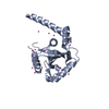


 PDBj
PDBj Assembly
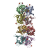


 Mass: 96.063 Da / Num. of mol.: 9 / Source method: obtained synthetically / Formula: SO4
 Mass: 92.094 Da / Num. of mol.: 10 / Source method: obtained synthetically / Formula: C3H8O3
 Mass: 35.453 Da / Num. of mol.: 4 / Source method: obtained synthetically / Formula: Cl
 Mass: 18.015 Da / Num. of mol.: 183 / Source method: isolated from a natural source / Formula: H2O
 Sample preparation
 / Beamline: 8.2.2 / Wavelength: 0.9537, 0.9798, 0.9796
 Processing