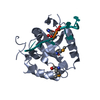
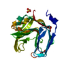
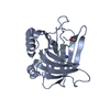
[English] 日本語
Yorodumi
- PDB-3wp5: The crystal structure of mutant CDBFV E109A from Neocallimastix p... -

+
Open data


ID or keywords:

Loading...

-
Basic information

Entry
Database: PDB / ID: 3wp5
TitleThe crystal structure of mutant CDBFV E109A from Neocallimastix patriciarum
ComponentsCDBFV
KeywordsHYDROLASE / xylanase / regulatory N-terminal region / disulfide bond / industrial enzyme / beta-jellyroll fold
Function / homology
Function and homology information


cellulose binding / endo-1,4-beta-xylanase activity / endo-1,4-beta-xylanase / xylan catabolic process / extracellular region
Similarity search - Function
CBM1 (carbohydrate binding type-1) domain signature. / Cellulose-binding domain, fungal / Cellulose-binding domain superfamily / Fungal cellulose binding domain / CBM1 (carbohydrate binding type-1) domain profile. / Fungal-type cellulose-binding domain / Glycoside hydrolase family 11/12, catalytic domain / Glycoside hydrolase family 11, active site 1 / Glycosyl hydrolases family 11 (GH11) active site signature 1. / Glycoside hydrolase family 11 ...CBM1 (carbohydrate binding type-1) domain signature. / Cellulose-binding domain, fungal / Cellulose-binding domain superfamily / Fungal cellulose binding domain / CBM1 (carbohydrate binding type-1) domain profile. / Fungal-type cellulose-binding domain / Glycoside hydrolase family 11/12, catalytic domain / Glycoside hydrolase family 11, active site 1 / Glycosyl hydrolases family 11 (GH11) active site signature 1. / Glycoside hydrolase family 11 / Glycosyl hydrolases family 11 (GH11) domain / Glycosyl hydrolases family 11 / Glycosyl hydrolases family 11 (GH11) domain profile. / Glycoside hydrolase family 11/12 / Concanavalin A-like lectin/glucanase domain superfamily / Jelly Rolls / Sandwich / Mainly Beta
Similarity search - Domain/homology
Endo-1,4-beta-xylanase C
Similarity search - Component
Biological speciesNeocallimastix patriciarum (fungus)
MethodX-RAY DIFFRACTION / SYNCHROTRON / MOLECULAR REPLACEMENT / Resolution: 1.32 Å
AuthorsCheng, Y.S. / Chen, C.C. / Huang, C.H. / Huang, T.Y. / Ko, T.P. / Huang, J.W. / Wu, T.H. / Liu, J.R. / Guo, R.T.
CitationJournal: J.Biol.Chem. / Year: 2014
Title: Structural analysis of a glycoside hydrolase family 11 xylanase from Neocallimastix patriciarum: insights into the molecular basis of a thermophilic enzyme.
Authors: Cheng, Y.S. / Chen, C.C. / Huang, C.H. / Ko, T.P. / Luo, W. / Huang, J.W. / Liu, J.R. / Guo, R.T.
History
DepositionJan 9, 2014Deposition site: PDBJ / Processing site: PDBJ
Revision 1.0Mar 19, 2014Provider: repository / Type: Initial release
Revision 1.1Dec 13, 2017Group: Database references / Category: pdbx_database_related / Item: _pdbx_database_related.db_name
Revision 1.2Dec 25, 2019Group: Database references / Category: citation
Item: _citation.journal_volume / _citation.page_first ..._citation.journal_volume / _citation.page_first / _citation.page_last / _citation.pdbx_database_id_PubMed / _citation.title
Revision 1.3Nov 8, 2023Group: Data collection / Database references / Refinement description
Category: chem_comp_atom / chem_comp_bond ...chem_comp_atom / chem_comp_bond / database_2 / pdbx_initial_refinement_model
Item: _database_2.pdbx_DOI / _database_2.pdbx_database_accession

-
Structure visualization

Structure viewerMolecule:
MolmilJmol/JSmol

Downloads & links

-
Assembly

Deposited unit
A: CDBFV


Theoretical massNumber of molelcules
Total (without water)24,6351
Polymers24,6351
Non-polymers00
Water6,323351
1


  • Idetical with deposited unit
  • defined by author&software
TypeNameSymmetry operationNumber
identity operation1_555x,y,z1
MethodPISA
Unit cell
Length a, b, c (Å)92.857, 92.857, 43.144
Angle α, β, γ (deg.)90.00, 90.00, 120.00
Int Tables number172
Space group name H-MP64

-
Components

#1: Protein CDBFV / xylanase


Mass: 24634.998 Da / Num. of mol.: 1
Source method: isolated from a genetically manipulated source
Source: (gene. exp.) Neocallimastix patriciarum (fungus) / Plasmid: pPICZalphaA / Production host: Komagataella pastoris (fungus) / References: UniProt: Q9UV68*PLUS
#2: Water ChemComp-HOH / water / Water


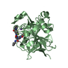
Mass: 18.015 Da / Num. of mol.: 351 / Source method: isolated from a natural source / Formula: H2O
Sequence detailsA SEQUENCE REFERENCE FOR THIS PROTEIN DOES NOT CURRENTLY EXIST. THIS PROTEIN HAS MUTATION E109A

-
Experimental details

-
Experiment

ExperimentMethod: X-RAY DIFFRACTION / Number of used crystals: 1

-
Sample preparation

CrystalDensity Matthews: 2.18 Å3/Da / Density % sol: 43.57 %
Crystal growTemperature: 298 K / Method: vapor diffusion, sitting drop / pH: 6.5
Details: 0.1M sodium cacodylate, pH 6.5, 0.2M ammonium sulfate, 26% PEG 8000, 5% glycerol, VAPOR DIFFUSION, SITTING DROP, temperature 298K

-
Data collection

DiffractionMean temperature: 100 K
Diffraction sourceSource: SYNCHROTRON / Site: NSRRC 
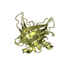
/ Beamline: BL13B1 / Wavelength: 1 Å
DetectorType: ADSC QUANTUM 4 / Detector: CCD / Date: Sep 13, 2012
RadiationProtocol: SINGLE WAVELENGTH / Monochromatic (M) / Laue (L): M / Scattering type: x-ray
Radiation wavelengthWavelength: 1 Å / Relative weight: 1
ReflectionResolution: 1.32→25 Å / Num. obs: 50174 / % possible obs: 100 % / Redundancy: 9.1 % / Rmerge(I) obs: 0.043 / Net I/σ(I): 44.8
Reflection shellResolution: 1.32→1.37 Å / Redundancy: 8.9 % / Rmerge(I) obs: 0.467 / Mean I/σ(I) obs: 3.1 / Num. unique all: 5000 / % possible all: 100

-
Processing

Software
NameVersionClassification
HKL-2000data collection
PHASERphasing
REFMAC5.7.0029refinement
HKL-2000data reduction
HKL-2000data scaling
RefinementMethod to determine structure: MOLECULAR REPLACEMENT
Starting model: 3WP4
Resolution: 1.32→24.86 Å / Cor.coef. Fo:Fc: 0.974 / Cor.coef. Fo:Fc free: 0.958 / SU B: 1.434 / SU ML: 0.028 / Cross valid method: THROUGHOUT / ESU R: 0.053 / ESU R Free: 0.051 / Stereochemistry target values: MAXIMUM LIKELIHOOD / Details: HYDROGENS HAVE BEEN USED IF PRESENT IN THE INPUT
RfactorNum. reflection% reflectionSelection details
Rfree0.18263 2544 5.1 %RANDOM
Rwork0.14673 ---
obs0.14851 47540 99.71 %-
Solvent computationIon probe radii: 0.8 Å / Shrinkage radii: 0.8 Å / VDW probe radii: 1.2 Å / Solvent model: MASK
Displacement parametersBiso mean: 14.931 Å2
Baniso -1Baniso -2Baniso -3
1-0.05 Å20.05 Å20 Å2
2--0.05 Å20 Å2
3----0.15 Å2
Refinement stepCycle: LAST / Resolution: 1.32→24.86 Å
ProteinNucleic acidLigandSolventTotal
Num. atoms1738 0 0 351 2089
Refine LS restraints
Refine-IDTypeDev idealDev ideal targetNumber
X-RAY DIFFRACTIONr_bond_refined_d0.0140.0191785
X-RAY DIFFRACTIONr_bond_other_d
X-RAY DIFFRACTIONr_angle_refined_deg1.7381.9172419
X-RAY DIFFRACTIONr_angle_other_deg
X-RAY DIFFRACTIONr_dihedral_angle_1_deg6.9165225
X-RAY DIFFRACTIONr_dihedral_angle_2_deg32.49824.40584
X-RAY DIFFRACTIONr_dihedral_angle_3_deg10.48215265
X-RAY DIFFRACTIONr_dihedral_angle_4_deg12.805156
X-RAY DIFFRACTIONr_chiral_restr0.1170.2247
X-RAY DIFFRACTIONr_gen_planes_refined0.010.0211403
X-RAY DIFFRACTIONr_gen_planes_other
X-RAY DIFFRACTIONr_nbd_refined
X-RAY DIFFRACTIONr_nbd_other
X-RAY DIFFRACTIONr_nbtor_refined
X-RAY DIFFRACTIONr_nbtor_other
X-RAY DIFFRACTIONr_xyhbond_nbd_refined
X-RAY DIFFRACTIONr_xyhbond_nbd_other
X-RAY DIFFRACTIONr_metal_ion_refined
X-RAY DIFFRACTIONr_metal_ion_other
X-RAY DIFFRACTIONr_symmetry_vdw_refined
X-RAY DIFFRACTIONr_symmetry_vdw_other
X-RAY DIFFRACTIONr_symmetry_hbond_refined
X-RAY DIFFRACTIONr_symmetry_hbond_other
X-RAY DIFFRACTIONr_symmetry_metal_ion_refined
X-RAY DIFFRACTIONr_symmetry_metal_ion_other
X-RAY DIFFRACTIONr_mcbond_it
X-RAY DIFFRACTIONr_mcbond_other
X-RAY DIFFRACTIONr_mcangle_it
X-RAY DIFFRACTIONr_mcangle_other
X-RAY DIFFRACTIONr_scbond_it
X-RAY DIFFRACTIONr_scbond_other
X-RAY DIFFRACTIONr_scangle_it
X-RAY DIFFRACTIONr_scangle_other
X-RAY DIFFRACTIONr_long_range_B_refined
X-RAY DIFFRACTIONr_long_range_B_other
X-RAY DIFFRACTIONr_rigid_bond_restr4.65631785
X-RAY DIFFRACTIONr_sphericity_free30.023596
X-RAY DIFFRACTIONr_sphericity_bonded7.68851993
LS refinement shellResolution: 1.318→1.353 Å / Total num. of bins used: 20
RfactorNum. reflection% reflection
Rfree0.254 181 -
Rwork0.212 3465 -
obs--98.59 %
Refinement TLS params.Method: refined / Origin x: -0.8588 Å / Origin y: -32.048 Å / Origin z: -4.8069 Å
111213212223313233
T0.012 Å20.0107 Å2-0.0066 Å2-0.0141 Å2-0.0028 Å2--0.0137 Å2
L0.596 °2-0.019 °2-0.144 °2-0.36 °2-0.1604 °2--0.2332 °2
S0.0451 Å °0.0304 Å °-0.0229 Å °-0.0277 Å °-0.0553 Å °-0.0058 Å °0.001 Å °0.0302 Å °0.0102 Å °

+
About Yorodumi

-
News

-
Feb 9, 2022. New format data for meta-information of EMDB entries

New format data for meta-information of EMDB entries

  • Version 3 of the EMDB header file is now the official format.
  • The previous official version 1.9 will be removed from the archive.

Related info.:EMDB header

External links:wwPDB to switch to version 3 of the EMDB data model

-
Aug 12, 2020. Covid-19 info

Covid-19 info

URL: https://pdbj.org/emnavi/covid19.php

New page: Covid-19 featured information page in EM Navigator.

Related info.:Covid-19 info / Mar 5, 2020. Novel coronavirus structure data

+
Mar 5, 2020. Novel coronavirus structure data

Novel coronavirus structure data

Related info.:Yorodumi Speices / Aug 12, 2020. Covid-19 info

External links:COVID-19 featured content - PDBj / Molecule of the Month (242):Coronavirus Proteases

+
Jan 31, 2019. EMDB accession codes are about to change! (news from PDBe EMDB page)

EMDB accession codes are about to change! (news from PDBe EMDB page)

  • The allocation of 4 digits for EMDB accession codes will soon come to an end. Whilst these codes will remain in use, new EMDB accession codes will include an additional digit and will expand incrementally as the available range of codes is exhausted. The current 4-digit format prefixed with “EMD-” (i.e. EMD-XXXX) will advance to a 5-digit format (i.e. EMD-XXXXX), and so on. It is currently estimated that the 4-digit codes will be depleted around Spring 2019, at which point the 5-digit format will come into force.
  • The EM Navigator/Yorodumi systems omit the EMD- prefix.

Related info.:Q: What is EMD? / ID/Accession-code notation in Yorodumi/EM Navigator

External links:EMDB Accession Codes are Changing Soon! / Contact to PDBj

+
Jul 12, 2017. Major update of PDB

Major update of PDB

  • wwPDB released updated PDB data conforming to the new PDBx/mmCIF dictionary.
  • This is a major update changing the version number from 4 to 5, and with Remediation, in which all the entries are updated.
  • In this update, many items about electron microscopy experimental information are reorganized (e.g. em_software).
  • Now, EM Navigator and Yorodumi are based on the updated data.

External links:wwPDB Remediation / Enriched Model Files Conforming to OneDep Data Standards Now Available in the PDB FTP Archive

-
Yorodumi

Thousand views of thousand structures

  • Yorodumi is a browser for structure data from EMDB, PDB, SASBDB, etc.
  • This page is also the successor to EM Navigator detail page, and also detail information page/front-end page for Omokage search.
  • The word "yorodu" (or yorozu) is an old Japanese word meaning "ten thousand". "mi" (miru) is to see.

Related info.:EMDB / PDB / SASBDB / Comparison of 3 databanks / Yorodumi Search / Aug 31, 2016. New EM Navigator & Yorodumi / Yorodumi Papers / Jmol/JSmol / Function and homology information / Changes in new EM Navigator and Yorodumi

Read more