 Movie
Movie Controller

[English] 日本語
 Yorodumi
- PDB-3wfe: Reduced and cyanide-bound cytochrome c-dependent nitric oxide red... -
+ Open data

- Basic information
| Entry | Database: PDB / ID: 3wfe | ||||||
|---|---|---|---|---|---|---|---|
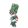
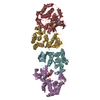
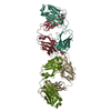
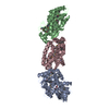
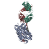
| Title | Reduced and cyanide-bound cytochrome c-dependent nitric oxide reductase (cNOR) from Pseudomonas aeruginosa in complex with antibody fragment | ||||||
 Components Components |
| ||||||
 Keywords Keywords | IMMUNE SYSTEM/OXIDOREDUCTASE / metal-binding / membrane protein / IMMUNE SYSTEM-OXIDOREDUCTASE complex | ||||||
| Function / homology |  Function and homology information Function and homology informationnitric oxide reductase (cytochrome c) / nitric oxide reductase activity / denitrification pathway / cytochrome-c oxidase activity / aerobic respiration / respiratory electron transport chain / electron transfer activity / heme binding / metal ion binding / plasma membrane Similarity search - Function | ||||||
| Biological species |    Pseudomonas aeruginosa (bacteria) Pseudomonas aeruginosa (bacteria) | ||||||
| Method |  X-RAY DIFFRACTION / SYNCHROTRON / MOLECULAR REPLACEMENT / Resolution: 2.49 Å X-RAY DIFFRACTION / SYNCHROTRON / MOLECULAR REPLACEMENT / Resolution: 2.49 Å | ||||||
 Authors Authors | Sato, N. / Ishii, S. / Hino, T. / Sugimoto, H. / Fukumori, Y. / Shiro, Y. / Tosha, T. | ||||||
 Citation Citation | Journal: Proteins / Year: 2014 Title: Structures of reduced and ligand-bound nitric oxide reductase provide insights into functional differences in respiratory enzymes. Authors: Sato, N. / Ishii, S. / Sugimoto, H. / Hino, T. / Fukumori, Y. / Sako, Y. / Shiro, Y. / Tosha, T. #1: Journal: Science / Year: 2010Title: Structural basis of biological N2O generation by bacterial nitric oxide reductase Authors: Hino, T. / Matsumoto, Y. / Nagano, S. / Sugimoto, H. / Fukumori, Y. / Murata, T. / Iwata, S. / Shiro, Y. | ||||||
| History |
|
- Structure visualization
| Structure viewer | Molecule: MolmilJmol/JSmol |
|---|
- Downloads & links
-Download
| PDBx/mmCIF format | 3wfe.cif.gz | 430.5 KB | Display | PDBx/mmCIF format |
|---|---|---|---|---|
| PDB format | pdb3wfe.ent.gz | 351.8 KB | Display | PDB format |
| PDBx/mmJSON format | 3wfe.json.gz | Tree view | PDBx/mmJSON format | |
| Others |  Other downloads Other downloads |
-Validation report
| Arichive directory | https://data.pdbj.org/pub/pdb/validation_reports/wf/3wfeftp://data.pdbj.org/pub/pdb/validation_reports/wf/3wfe | HTTPS FTP |
|---|
-Related structure data
| Related structure data | 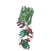 3wfbC  3wfcC  3wfdC  3o0rS S: Starting model for refinement C: citing same article ( |
|---|---|
| Similar structure data |


-Links
 PDBj
PDBj


- Assembly
| Deposited unit | 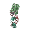
| ||||||||
|---|---|---|---|---|---|---|---|---|---|
| 1 |
| ||||||||
| Unit cell |
|
-Components
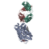
-Nitric oxide reductase subunit ... , 2 types, 2 molecules BC
| #3: Protein | Mass: 52215.871 Da / Num. of mol.: 1 / Source method: isolated from a natural source / Source: (natural) Pseudomonas aeruginosa (bacteria) / Strain: PAO1References: UniProt: Q59647, nitric oxide reductase (cytochrome c) |
|---|---|
| #4: Protein | Mass: 16374.622 Da / Num. of mol.: 1 / Source method: isolated from a natural source / Source: (natural) Pseudomonas aeruginosa (bacteria) / Strain: PAO1 / References: UniProt: Q59646 |
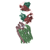
-Antibody , 2 types, 2 molecules LH
| #1: Antibody | Mass: 23735.326 Da / Num. of mol.: 1 / Source method: isolated from a natural source / Source: (natural) |
|---|---|
| #2: Antibody | Mass: 24089.945 Da / Num. of mol.: 1 / Source method: isolated from a natural source / Source: (natural) |
-Sugars , 1 types, 2 molecules 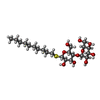
| #8: Sugar |  Type: D-saccharide / Mass: 498.628 Da / Num. of mol.: 2 Type: D-saccharide / Mass: 498.628 Da / Num. of mol.: 2Source method: isolated from a genetically manipulated source Formula: C22H42O10S / Comment: detergent*YM |
|---|
-Non-polymers , 6 types, 249 molecules 


| #5: Chemical |  Mass: 616.487 Da / Num. of mol.: 2 / Source method: obtained synthetically / Formula: C34H32FeN4O4 Mass: 616.487 Da / Num. of mol.: 2 / Source method: obtained synthetically / Formula: C34H32FeN4O4#6: Chemical | ChemComp-FE / |  Mass: 55.845 Da / Num. of mol.: 1 / Source method: obtained synthetically / Formula: Fe Mass: 55.845 Da / Num. of mol.: 1 / Source method: obtained synthetically / Formula: Fe#7: Chemical |  Mass: 26.017 Da / Num. of mol.: 2 / Source method: obtained synthetically / Formula: CN Mass: 26.017 Da / Num. of mol.: 2 / Source method: obtained synthetically / Formula: CN#9: Chemical | ChemComp-CA / |  Mass: 40.078 Da / Num. of mol.: 1 / Source method: obtained synthetically / Formula: Ca Mass: 40.078 Da / Num. of mol.: 1 / Source method: obtained synthetically / Formula: Ca#10: Chemical | ChemComp-HEC / |  Mass: 618.503 Da / Num. of mol.: 1 / Source method: obtained synthetically / Formula: C34H34FeN4O4 Mass: 618.503 Da / Num. of mol.: 1 / Source method: obtained synthetically / Formula: C34H34FeN4O4#11: Water | ChemComp-HOH / | Mass: 18.015 Da / Num. of mol.: 242 / Source method: isolated from a natural source / Formula: H2O |
|---|
-Details
| Has protein modification | Y |
|---|
-Experimental details
-Experiment
| Experiment | Method: X-RAY DIFFRACTION / Number of used crystals: 1 |
|---|
- Sample preparation
| Crystal | Density Matthews: 4.08 Å3/Da / Density % sol: 69.88 % |
|---|---|
| Crystal grow | Temperature: 277 K / Method: vapor diffusion / pH: 6 Details: 100mM sodium citrate, pH 6.0, vapor diffusion, temperature 277K |
-Data collection
| Diffraction | Mean temperature: 100 K |
|---|---|
| Diffraction source | Source: SYNCHROTRON / Site: SPring-8  / Beamline: BL41XU / Wavelength: 1 Å / Beamline: BL41XU / Wavelength: 1 Å |
| Detector | Type: RAYONIX MX225HE / Detector: CCD / Date: Jan 24, 2012 / Details: mirrors |
| Diffraction measurement | Details: 0.60 degrees, 1.8 sec, detector distance 220.00 mm / Method: \w scans |
| Radiation | Monochromator: Si(111) / Protocol: SINGLE WAVELENGTH / Monochromatic (M) / Laue (L): M / Scattering type: x-ray |
| Radiation wavelength | Wavelength: 1 Å / Relative weight: 1 |
| Reflection | Av R equivalents: 0.076 / Number: 492828 |
| Reflection | Resolution: 2.49→50 Å / Num. obs: 66399 / % possible obs: 99.1 % / Observed criterion σ(F): 0 / Observed criterion σ(I): -3 / Redundancy: 7.4 % / Rmerge(I) obs: 0.076 / Rsym value: 0.076 / Net I/σ(I): 24.231 |
| Reflection shell | Resolution: 2.5→2.59 Å / Redundancy: 7.5 % / Rmerge(I) obs: 0.526 / Mean I/σ(I) obs: 3.684 / Rsym value: 0.526 / % possible all: 100 |
| Cell measurement | Reflection used: 492828 |
- Processing
| Software |
| ||||||||||||||||||||||||||||||||||||||||||||||||||||||||||||||||||||||||||||||||||||||||||||||||||||||||||||||||||||||||||||||||||||||||||||||||||||||||||||||||||||||||||||||||||||||||||||||||||||||||
|---|---|---|---|---|---|---|---|---|---|---|---|---|---|---|---|---|---|---|---|---|---|---|---|---|---|---|---|---|---|---|---|---|---|---|---|---|---|---|---|---|---|---|---|---|---|---|---|---|---|---|---|---|---|---|---|---|---|---|---|---|---|---|---|---|---|---|---|---|---|---|---|---|---|---|---|---|---|---|---|---|---|---|---|---|---|---|---|---|---|---|---|---|---|---|---|---|---|---|---|---|---|---|---|---|---|---|---|---|---|---|---|---|---|---|---|---|---|---|---|---|---|---|---|---|---|---|---|---|---|---|---|---|---|---|---|---|---|---|---|---|---|---|---|---|---|---|---|---|---|---|---|---|---|---|---|---|---|---|---|---|---|---|---|---|---|---|---|---|---|---|---|---|---|---|---|---|---|---|---|---|---|---|---|---|---|---|---|---|---|---|---|---|---|---|---|---|---|---|---|---|---|
| Refinement | Method to determine structure: MOLECULAR REPLACEMENT Starting model: 3O0R Resolution: 2.49→35.16 Å / Cor.coef. Fo:Fc: 0.951 / Cor.coef. Fo:Fc free: 0.934 / Occupancy max: 1 / Occupancy min: 1 / SU B: 12.756 / SU ML: 0.146 / Cross valid method: THROUGHOUT / σ(F): 0 / ESU R: 0.262 / ESU R Free: 0.214 / Stereochemistry target values: MAXIMUM LIKELIHOOD Details: HYDROGENS HAVE BEEN ADDED IN THE RIDING POSITIONS U VALUES: WITH TLS ADDED
| ||||||||||||||||||||||||||||||||||||||||||||||||||||||||||||||||||||||||||||||||||||||||||||||||||||||||||||||||||||||||||||||||||||||||||||||||||||||||||||||||||||||||||||||||||||||||||||||||||||||||
| Solvent computation | Ion probe radii: 0.8 Å / Shrinkage radii: 0.8 Å / VDW probe radii: 1.2 Å / Solvent model: MASK | ||||||||||||||||||||||||||||||||||||||||||||||||||||||||||||||||||||||||||||||||||||||||||||||||||||||||||||||||||||||||||||||||||||||||||||||||||||||||||||||||||||||||||||||||||||||||||||||||||||||||
| Displacement parameters | Biso max: 170.95 Å2 / Biso mean: 61.6746 Å2 / Biso min: 33.48 Å2
| ||||||||||||||||||||||||||||||||||||||||||||||||||||||||||||||||||||||||||||||||||||||||||||||||||||||||||||||||||||||||||||||||||||||||||||||||||||||||||||||||||||||||||||||||||||||||||||||||||||||||
| Refinement step | Cycle: LAST / Resolution: 2.49→35.16 Å
| ||||||||||||||||||||||||||||||||||||||||||||||||||||||||||||||||||||||||||||||||||||||||||||||||||||||||||||||||||||||||||||||||||||||||||||||||||||||||||||||||||||||||||||||||||||||||||||||||||||||||
| Refine LS restraints |
| ||||||||||||||||||||||||||||||||||||||||||||||||||||||||||||||||||||||||||||||||||||||||||||||||||||||||||||||||||||||||||||||||||||||||||||||||||||||||||||||||||||||||||||||||||||||||||||||||||||||||
| LS refinement shell | Resolution: 2.494→2.559 Å / Total num. of bins used: 20
| ||||||||||||||||||||||||||||||||||||||||||||||||||||||||||||||||||||||||||||||||||||||||||||||||||||||||||||||||||||||||||||||||||||||||||||||||||||||||||||||||||||||||||||||||||||||||||||||||||||||||
| Refinement TLS params. | Method: refined / Refine-ID: X-RAY DIFFRACTION
| ||||||||||||||||||||||||||||||||||||||||||||||||||||||||||||||||||||||||||||||||||||||||||||||||||||||||||||||||||||||||||||||||||||||||||||||||||||||||||||||||||||||||||||||||||||||||||||||||||||||||
| Refinement TLS group |
|
