 Movie
Movie Controller


+ Open data

- Basic information
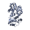
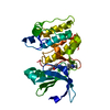
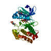
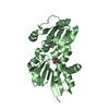
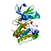
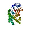
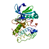
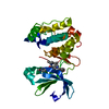
| Entry | Database: PDB / ID: 2f5x | ||||||
|---|---|---|---|---|---|---|---|
| Title | Structure of periplasmic binding protein BugD | ||||||
 Components Components | BugD | ||||||
 Keywords Keywords | TRANSPORT PROTEIN / periplasmic binding protein | ||||||
| Function / homology |  Function and homology information Function and homology informationBordetella uptake gene, domain 1 / Bordetella uptake gene / Bordetella uptake gene, domain 1 / Tripartite tricarboxylate transporter family receptor / Periplasmic binding protein-like II / D-Maltodextrin-Binding Protein; domain 2 / 3-Layer(aba) Sandwich / Alpha Beta Similarity search - Domain/homology | ||||||
| Biological species |  Bordetella pertussis Tohama I (bacteria) Bordetella pertussis Tohama I (bacteria) | ||||||
| Method |  X-RAY DIFFRACTION / SYNCHROTRON / SAD / Resolution: 1.72 Å X-RAY DIFFRACTION / SYNCHROTRON / SAD / Resolution: 1.72 Å | ||||||
 Authors Authors | Huvent, I. / Belrhali, H. / Antoine, R. / Bompard, C. / Jacob-Dubuisson, F. / Villeret, V. | ||||||
 Citation Citation | Journal: J.Mol.Biol. / Year: 2006 Title: Crystal Structure of Bordetella pertussis BugD Solute Receptor Unveils the Basis of Ligand Binding in a New Family of Periplasmic Binding Proteins Authors: Huvent, I. / Belrhali, H. / Antoine, R. / Bompard, C. / Locht, C. / Jacob-Dubuisson, F. / Villeret, V. | ||||||
| History |
|
- Structure visualization
| Structure viewer | Molecule: MolmilJmol/JSmol |
|---|
- Downloads & links
-Download
| PDBx/mmCIF format | 2f5x.cif.gz | 387.3 KB | Display | PDBx/mmCIF format |
|---|---|---|---|---|
| PDB format | pdb2f5x.ent.gz | 316.5 KB | Display | PDB format |
| PDBx/mmJSON format | 2f5x.json.gz | Tree view | PDBx/mmJSON format | |
| Others |  Other downloads Other downloads |
-Validation report
| Arichive directory | https://data.pdbj.org/pub/pdb/validation_reports/f5/2f5xftp://data.pdbj.org/pub/pdb/validation_reports/f5/2f5x | HTTPS FTP |
|---|
-Related structure data
| Similar structure data |
|---|


-Links
 PDBj
PDBj
- Assembly
| Deposited unit | 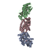
| ||||||||
|---|---|---|---|---|---|---|---|---|---|
| 1 | 
| ||||||||
| 2 | 
| ||||||||
| 3 | 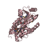
| ||||||||
| Unit cell |
| ||||||||
| Details | the biological assembly is one monomer. THe asymmetric unit contains three monomers |
-Components
| #1: Protein | Mass: 34030.348 Da / Num. of mol.: 3 / Fragment: residues 26-325 Source method: isolated from a genetically manipulated source Source: (gene. exp.) Bordetella pertussis Tohama I (bacteria)Species: Bordetella pertussis / Strain: Tohama I / Plasmid: pQE-30 / Production host: #2: Chemical |   Type: L-peptide linking / Mass: 133.103 Da / Num. of mol.: 3 / Source method: obtained synthetically / Formula: C4H7NO4 Type: L-peptide linking / Mass: 133.103 Da / Num. of mol.: 3 / Source method: obtained synthetically / Formula: C4H7NO4#3: Water | ChemComp-HOH / |  Mass: 18.015 Da / Num. of mol.: 1107 / Source method: isolated from a natural source / Formula: H2O Mass: 18.015 Da / Num. of mol.: 1107 / Source method: isolated from a natural source / Formula: H2OHas protein modification | Y | |
|---|
-Experimental details
-Experiment
| Experiment | Method: X-RAY DIFFRACTION / Number of used crystals: 1 |
|---|
- Sample preparation
| Crystal | Density Matthews: 2.43 Å3/Da / Density % sol: 49.29 % |
|---|---|
| Crystal grow | Temperature: 278 K / Method: vapor diffusion, hanging drop / pH: 8 Details: Tris 10 mM, (NH4)2SO4 0.2 M, PEG1500 36% (w/v), glycerol 15%, pH 8.00, VAPOR DIFFUSION, HANGING DROP, temperature 278K |
-Data collection
| Diffraction | Mean temperature: 100 K |
|---|---|
| Diffraction source | Source: SYNCHROTRON / Site: ESRF  / Beamline: BM14 / Wavelength: 0.9791 / Beamline: BM14 / Wavelength: 0.9791 |
| Detector | Type: MARMOSAIC 225 mm CCD / Detector: CCD / Date: Aug 8, 2004 |
| Radiation | Protocol: SINGLE WAVELENGTH / Monochromatic (M) / Laue (L): M / Scattering type: x-ray |
| Radiation wavelength | Wavelength: 0.9791 Å / Relative weight: 1 |
| Reflection | Resolution: 1.72→49.63 Å / Num. obs: 101925 / % possible obs: 99.8 % / Observed criterion σ(I): 2 / Redundancy: 10.9 % / Rsym value: 0.072 / Net I/σ(I): 7.1 |
| Reflection shell | Resolution: 1.72→1.81 Å / Redundancy: 10.8 % / Mean I/σ(I) obs: 3 / Rsym value: 0.243 / % possible all: 99.6 |
- Processing
| Software |
| ||||||||||||||||||||||||||||||||||||||||||||||||||||||||||||||||||||||||||||||||||||||||||||||||||||||||||||||||||||||||||||||||||||||||||||||||||||||||||||||||||||||||||
|---|---|---|---|---|---|---|---|---|---|---|---|---|---|---|---|---|---|---|---|---|---|---|---|---|---|---|---|---|---|---|---|---|---|---|---|---|---|---|---|---|---|---|---|---|---|---|---|---|---|---|---|---|---|---|---|---|---|---|---|---|---|---|---|---|---|---|---|---|---|---|---|---|---|---|---|---|---|---|---|---|---|---|---|---|---|---|---|---|---|---|---|---|---|---|---|---|---|---|---|---|---|---|---|---|---|---|---|---|---|---|---|---|---|---|---|---|---|---|---|---|---|---|---|---|---|---|---|---|---|---|---|---|---|---|---|---|---|---|---|---|---|---|---|---|---|---|---|---|---|---|---|---|---|---|---|---|---|---|---|---|---|---|---|---|---|---|---|---|---|---|---|
| Refinement | Method to determine structure: SAD / Resolution: 1.72→49.63 Å / Cor.coef. Fo:Fc: 0.965 / Cor.coef. Fo:Fc free: 0.953 / SU B: 1.382 / SU ML: 0.047 / Cross valid method: THROUGHOUT / σ(F): 0 / ESU R: 0.135 / ESU R Free: 0.087 / Stereochemistry target values: MAXIMUM LIKELIHOOD / Details: HYDROGENS HAVE BEEN ADDED IN THE RIDING POSITIONS
| ||||||||||||||||||||||||||||||||||||||||||||||||||||||||||||||||||||||||||||||||||||||||||||||||||||||||||||||||||||||||||||||||||||||||||||||||||||||||||||||||||||||||||
| Solvent computation | Ion probe radii: 0.8 Å / Shrinkage radii: 0.8 Å / VDW probe radii: 1.4 Å / Solvent model: MASK | ||||||||||||||||||||||||||||||||||||||||||||||||||||||||||||||||||||||||||||||||||||||||||||||||||||||||||||||||||||||||||||||||||||||||||||||||||||||||||||||||||||||||||
| Displacement parameters | Biso mean: 16.49 Å2
| ||||||||||||||||||||||||||||||||||||||||||||||||||||||||||||||||||||||||||||||||||||||||||||||||||||||||||||||||||||||||||||||||||||||||||||||||||||||||||||||||||||||||||
| Refinement step | Cycle: LAST / Resolution: 1.72→49.63 Å
| ||||||||||||||||||||||||||||||||||||||||||||||||||||||||||||||||||||||||||||||||||||||||||||||||||||||||||||||||||||||||||||||||||||||||||||||||||||||||||||||||||||||||||
| Refine LS restraints |
| ||||||||||||||||||||||||||||||||||||||||||||||||||||||||||||||||||||||||||||||||||||||||||||||||||||||||||||||||||||||||||||||||||||||||||||||||||||||||||||||||||||||||||
| LS refinement shell | Resolution: 1.72→1.76 Å / Total num. of bins used: 20 /
| ||||||||||||||||||||||||||||||||||||||||||||||||||||||||||||||||||||||||||||||||||||||||||||||||||||||||||||||||||||||||||||||||||||||||||||||||||||||||||||||||||||||||||
| Refinement TLS params. | Method: refined / Origin x: 15.133 Å / Origin y: 36.774 Å / Origin z: 1.436 Å
| ||||||||||||||||||||||||||||||||||||||||||||||||||||||||||||||||||||||||||||||||||||||||||||||||||||||||||||||||||||||||||||||||||||||||||||||||||||||||||||||||||||||||||
| Refinement TLS group |
|
