- PDB-3w5a: Crystal structure of the calcium pump and sarcolipin from rabbit ... -
+
Open data
ID or keywords:
Loading...
-
Basic information
Entry
Database: PDB / ID: 3w5a
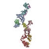
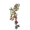
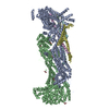
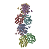
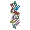
Title
Crystal structure of the calcium pump and sarcolipin from rabbit fast twitch skeletal muscle in the E1.Mg2+ state
Components
SERCA1a
Sarcolipin
Keywords
METAL TRANSPORT/MEMBRANE PROTEIN / P-TYPE ATPASE / HYDROLASE / CALCIUM TRANSPORT / CALCIUM BINDING / ATP BINDING / ENDOPLASMIC RETICULUM / SARCOLIPIN / METAL TRANSPORT-MEMBRANE PROTEIN complex
Function / homology
Function and homology information
positive regulation of protein depolymerization / regulation of ATPase-coupled calcium transmembrane transporter activity / regulation of relaxation of muscle / negative regulation of calcium ion import into sarcoplasmic reticulum / negative regulation of ATPase-coupled calcium transmembrane transporter activity / negative regulation of calcium ion import / negative regulation of protein-containing complex disassembly / positive regulation of calcium ion import into sarcoplasmic reticulum / positive regulation of ATPase-coupled calcium transmembrane transporter activity / positive regulation of fast-twitch skeletal muscle fiber contraction ...positive regulation of protein depolymerization / regulation of ATPase-coupled calcium transmembrane transporter activity / regulation of relaxation of muscle / negative regulation of calcium ion import into sarcoplasmic reticulum / negative regulation of ATPase-coupled calcium transmembrane transporter activity / negative regulation of calcium ion import / negative regulation of protein-containing complex disassembly / positive regulation of calcium ion import into sarcoplasmic reticulum / positive regulation of ATPase-coupled calcium transmembrane transporter activity / positive regulation of fast-twitch skeletal muscle fiber contraction / H zone / calcium ion import into sarcoplasmic reticulum / negative regulation of striated muscle contraction / regulation of striated muscle contraction / sarcoplasmic reticulum calcium ion transport / P-type Ca2+ transporter / P-type calcium transporter activity / positive regulation of cardiac muscle cell contraction / regulation of cardiac muscle contraction by calcium ion signaling / I band / endoplasmic reticulum-Golgi intermediate compartment / regulation of calcium ion transport / sarcoplasmic reticulum membrane / enzyme inhibitor activity / sarcoplasmic reticulum / calcium ion transport / ATPase binding / calcium ion binding / endoplasmic reticulum membrane / perinuclear region of cytoplasm / endoplasmic reticulum / ATP hydrolysis activity / ATP binding / membrane / metal ion binding Similarity search - Function
Resolution: 3→3.1 Å / Redundancy: 2.4 % / Rsym value: 0.412 / % possible all: 79.3
-
Processing
Software
Name
Version
Classification
BSS
datacollection
CNS
refinement
REFMAC
5.6.0117
refinement
DENZO
datareduction
SCALEPACK
datascaling
CNS
phasing
Refinement
Method to determine structure: MOLECULAR REPLACEMENT / Resolution: 3.01→15 Å / Cor.coef. Fo:Fc: 0.906 / Cor.coef. Fo:Fc free: 0.889 / SU B: 23.856 / SU ML: 0.406 / Cross valid method: THROUGHOUT / ESU R Free: 0.457 / Stereochemistry target values: MAXIMUM LIKELIHOOD / Details: HYDROGENS HAVE BEEN USED IF PRESENT IN THE INPUT
Rfactor
Num. reflection
% reflection
Selection details
Rfree
0.28307
3113
5 %
RANDOM
Rwork
0.24764
-
-
-
all
0.24945
-
-
-
obs
0.24945
58572
95.12 %
-
Solvent computation
Ion probe radii: 0.8 Å / Shrinkage radii: 0.8 Å / VDW probe radii: 1.2 Å / Solvent model: MASK
Displacement parameters
Biso mean: 78.777 Å2
Baniso -1
Baniso -2
Baniso -3
1-
2.89 Å2
0 Å2
3.98 Å2
2-
-
-0.41 Å2
0 Å2
3-
-
-
1.38 Å2
Refinement step
Cycle: LAST / Resolution: 3.01→15 Å
Protein
Nucleic acid
Ligand
Solvent
Total
Num. atoms
15613
0
177
12
15802
Refine LS restraints
Refine-ID
Type
Dev ideal
Dev ideal target
Number
X-RAY DIFFRACTION
r_bond_refined_d
0.007
0.019
16077
X-RAY DIFFRACTION
r_bond_other_d
X-RAY DIFFRACTION
r_angle_refined_deg
1.272
1.985
21817
X-RAY DIFFRACTION
r_angle_other_deg
X-RAY DIFFRACTION
r_dihedral_angle_1_deg
4.541
5
2016
X-RAY DIFFRACTION
r_dihedral_angle_2_deg
41.62
24.299
649
X-RAY DIFFRACTION
r_dihedral_angle_3_deg
18.239
15
2815
X-RAY DIFFRACTION
r_dihedral_angle_4_deg
15.489
15
99
X-RAY DIFFRACTION
r_chiral_restr
0.072
0.2
2532
X-RAY DIFFRACTION
r_gen_planes_refined
0.004
0.021
11866
X-RAY DIFFRACTION
r_gen_planes_other
X-RAY DIFFRACTION
r_nbd_refined
X-RAY DIFFRACTION
r_nbd_other
X-RAY DIFFRACTION
r_nbtor_refined
X-RAY DIFFRACTION
r_nbtor_other
X-RAY DIFFRACTION
r_xyhbond_nbd_refined
X-RAY DIFFRACTION
r_xyhbond_nbd_other
X-RAY DIFFRACTION
r_metal_ion_refined
X-RAY DIFFRACTION
r_metal_ion_other
X-RAY DIFFRACTION
r_symmetry_vdw_refined
X-RAY DIFFRACTION
r_symmetry_vdw_other
X-RAY DIFFRACTION
r_symmetry_hbond_refined
X-RAY DIFFRACTION
r_symmetry_hbond_other
X-RAY DIFFRACTION
r_symmetry_metal_ion_refined
X-RAY DIFFRACTION
r_symmetry_metal_ion_other
X-RAY DIFFRACTION
r_mcbond_it
X-RAY DIFFRACTION
r_mcbond_other
X-RAY DIFFRACTION
r_mcangle_it
X-RAY DIFFRACTION
r_scbond_it
X-RAY DIFFRACTION
r_scangle_it
X-RAY DIFFRACTION
r_rigid_bond_restr
X-RAY DIFFRACTION
r_sphericity_free
X-RAY DIFFRACTION
r_sphericity_bonded
LS refinement shell
Resolution: 3.006→3.081 Å / Total num. of bins used: 20
Rfactor
Num. reflection
% reflection
Rfree
0.425
180
-
Rwork
0.396
3211
-
obs
-
-
76.07 %
+
About Yorodumi
-
News
-
Feb 9, 2022. New format data for meta-information of EMDB entries
New format data for meta-information of EMDB entries
Version 3 of the EMDB header file is now the official format.
The previous official version 1.9 will be removed from the archive.
In the structure databanks used in Yorodumi, some data are registered as the other names, "COVID-19 virus" and "2019-nCoV". Here are the details of the virus and the list of structure data.
Jan 31, 2019. EMDB accession codes are about to change! (news from PDBe EMDB page)
EMDB accession codes are about to change! (news from PDBe EMDB page)
The allocation of 4 digits for EMDB accession codes will soon come to an end. Whilst these codes will remain in use, new EMDB accession codes will include an additional digit and will expand incrementally as the available range of codes is exhausted. The current 4-digit format prefixed with “EMD-” (i.e. EMD-XXXX) will advance to a 5-digit format (i.e. EMD-XXXXX), and so on. It is currently estimated that the 4-digit codes will be depleted around Spring 2019, at which point the 5-digit format will come into force.
The EM Navigator/Yorodumi systems omit the EMD- prefix.
Related info.:Q: What is EMD? / ID/Accession-code notation in Yorodumi/EM Navigator
Yorodumi is a browser for structure data from EMDB, PDB, SASBDB, etc.
This page is also the successor to EM Navigator detail page, and also detail information page/front-end page for Omokage search.
The word "yorodu" (or yorozu) is an old Japanese word meaning "ten thousand". "mi" (miru) is to see.
Related info.:EMDB / PDB / SASBDB / Comparison of 3 databanks / Yorodumi Search / Aug 31, 2016. New EM Navigator & Yorodumi / Yorodumi Papers / Jmol/JSmol / Function and homology information / Changes in new EM Navigator and Yorodumi
 Movie
Movie Controller

 Yorodumi
 Open data
 Basic information
 Components
 Keywords
 Function and homology information
 X-RAY DIFFRACTION /
  Authors
 Citation
 Structure visualization
 Downloads & links
 Other downloads
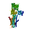


 PDBj
PDBj

 Assembly
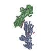
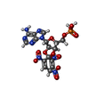


 Mass: 558.310 Da / Num. of mol.: 2 / Source method: obtained synthetically / Formula: C16H15N8O13P
 Mass: 734.039 Da / Num. of mol.: 5 / Source method: obtained synthetically / Formula: C40H80NO8P / Comment: phospholipid*YM
 Mass: 24.305 Da / Num. of mol.: 4 / Source method: obtained synthetically / Formula: Mg
 Mass: 22.990 Da / Num. of mol.: 2 / Source method: obtained synthetically / Formula: Na
 Sample preparation
 / Beamline: BL41XU / Wavelength: 0.9 Å
 Processing