 Movie
Movie Controller


+ Open data

- Basic information
| Entry | Database: PDB / ID: 3w3d | |||||||||
|---|---|---|---|---|---|---|---|---|---|---|
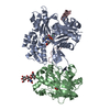
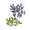
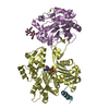
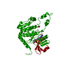
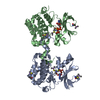
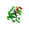
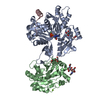
| Title | Crystal structure of smooth muscle G actin DNase I complex | |||||||||
 Components Components |
| |||||||||
 Keywords Keywords | STRUCTURAL PROTEIN / smooth muscle actin / actin / DNase I / G-actin / ATP Binding | |||||||||
| Function / homology |  Function and homology information Function and homology informationregulation of neutrophil mediated cytotoxicity / zymogen granule / Smooth Muscle Contraction / regulation of acute inflammatory response / deoxyribonuclease I / Regulation of CDH1 Function / neutrophil activation involved in immune response / deoxyribonuclease I activity / DNA catabolic process / cell periphery ...regulation of neutrophil mediated cytotoxicity / zymogen granule / Smooth Muscle Contraction / regulation of acute inflammatory response / deoxyribonuclease I / Regulation of CDH1 Function / neutrophil activation involved in immune response / deoxyribonuclease I activity / DNA catabolic process / cell periphery / Hydrolases; Acting on acid anhydrides; Acting on acid anhydrides to facilitate cellular and subcellular movement / nuclear envelope / actin cytoskeleton / actin binding / hydrolase activity / apoptotic process / DNA binding / extracellular region / ATP binding / nucleus Similarity search - Function | |||||||||
| Biological species |   | |||||||||
| Method |  X-RAY DIFFRACTION / SYNCHROTRON / MIR / Resolution: 1.8 Å X-RAY DIFFRACTION / SYNCHROTRON / MIR / Resolution: 1.8 Å | |||||||||
 Authors Authors | Sakabe, N. / Sakabe, K. / Sasaki, K. / Kondo, H. / Shimomur, M. | |||||||||
 Citation Citation | Journal: Acta Crystallogr.,Sect.A / Year: 1993 Title: Refined structure and solvent network of chicken gizzard G-actin DNase 1 complex at 1.8A resolution Authors: Sasaki, K. / Sakabe, K. / Sakabe, N. / Kondo, H. / Shimomur, M. #1: Journal: Nucl Instrum Methods Phys Res A / Year: 1991Title: X-ray diffraction data collection system for modern protein crystallography with a Weissenberg camera and an imaging plate using synchrotron radiation Authors: Sakabe, N. #2: Journal: J.Biochem. / Year: 1984 Title: X-ray diffraction photographs of chicken gizzard G-actin.DNase I complex crystals taken with synchrotron radiation. Authors: Sakabe, N. / Kamiya, N. / Sakabe, K. / Kondo, H. #3: Journal: J.Appl.Crystallogr. / Year: 1983Title: A F ocusing Weissenberg Camera with Multi-Layer-Line Screens for Macromolecular C rystallography Authors: Sakabe, N. #4: Journal: J.Biochem. / Year: 1983 Title: Crystallographic studies of the chicken gizzard G-actin X DNase I complex at 5A resolution Authors: Sakabe, N. / Sakabe, K. / Sasaki, K. / Kondo, H. / Ema, T. / Kamiya, N. / Matsushima, M. #5: Journal: J.Biochem. / Year: 1979 Title: Crystallization and preliminary crystallographic data of chicken gizzard G-actin . DNase I complex and Physarum G-actin . DNase I complex Authors: Sugino, H. / Sakabe, N. / Sakabe, K. / Hatano, S. / Oosawa, F. / Mikawa, T. / Ebashi, S. #6: Journal: Febs Lett. / Year: 1979 Title: The amino acid sequence of actin from chicken skeletal muscle actin and chicken gizzard smooth muscle actin Authors: Vandekerckhove, J. / Weber, K. | |||||||||
| History |
|
- Structure visualization
| Structure viewer | Molecule: MolmilJmol/JSmol |
|---|
- Downloads & links
-Download
| PDBx/mmCIF format | 3w3d.cif.gz | 154.4 KB | Display | PDBx/mmCIF format |
|---|---|---|---|---|
| PDB format | pdb3w3d.ent.gz | 117.2 KB | Display | PDB format |
| PDBx/mmJSON format | 3w3d.json.gz | Tree view | PDBx/mmJSON format | |
| Others |  Other downloads Other downloads |
-Validation report
| Arichive directory | https://data.pdbj.org/pub/pdb/validation_reports/w3/3w3dftp://data.pdbj.org/pub/pdb/validation_reports/w3/3w3d | HTTPS FTP |
|---|
-Related structure data
| Similar structure data |
|---|


-Links
 PDBj
PDBj


- Assembly
| Deposited unit | 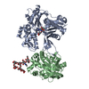
| ||||||||
|---|---|---|---|---|---|---|---|---|---|
| 1 |
| ||||||||
| Unit cell |
|
-Components
-Protein , 2 types, 2 molecules AB
| #1: Protein | Mass: 41732.492 Da / Num. of mol.: 1 / Source method: isolated from a natural source / Details: Gizzard / Source: (natural) |
|---|---|
| #2: Protein | Mass: 29092.574 Da / Num. of mol.: 1 / Source method: isolated from a natural source / Details: pancreas / Source: (natural) |
-Sugars , 1 types, 1 molecules
| #3: Polysaccharide | alpha-D-mannopyranose-(1-6)-beta-D-mannopyranose-(1-3)-[beta-D-mannopyranose-(1-3)-alpha-D- ...alpha-D-mannopyranose-(1-6)-beta-D-mannopyranose-(1-3)-[beta-D-mannopyranose-(1-3)-alpha-D-mannopyranose-(1-6)]beta-D-mannopyranose-(1-4)-2-acetamido-2-deoxy-beta-D-glucopyranose-(1-4)-2-acetamido-2-deoxy-beta-D-glucopyranose Source method: isolated from a genetically manipulated source |
|---|
-Non-polymers , 3 types, 386 molecules 


| #4: Chemical | ChemComp-ATP /  Mass: 507.181 Da / Num. of mol.: 1 / Source method: obtained synthetically / Formula: C10H16N5O13P3 / Comment: ATP, energy-carrying molecule*YM Mass: 507.181 Da / Num. of mol.: 1 / Source method: obtained synthetically / Formula: C10H16N5O13P3 / Comment: ATP, energy-carrying molecule*YM | ||
|---|---|---|---|
| #5: Chemical |  Mass: 40.078 Da / Num. of mol.: 2 / Source method: obtained synthetically / Formula: Ca Mass: 40.078 Da / Num. of mol.: 2 / Source method: obtained synthetically / Formula: Ca#6: Water | ChemComp-HOH / | Mass: 18.015 Da / Num. of mol.: 383 / Source method: isolated from a natural source / Formula: H2O |
-Details
| Has protein modification | Y |
|---|
-Experimental details
-Experiment
| Experiment | Method: X-RAY DIFFRACTION / Number of used crystals: 5 |
|---|
- Sample preparation
| Crystal | Density Matthews: 2.59 Å3/Da / Density % sol: 52.42 % |
|---|---|
| Crystal grow | Temperature: 300 K / Method: precipitation / pH: 6.6 Details: 4mM MgCl2, 0.1mM CaCl2, NaN3, PEG 6000, pH 6.6, precipitation , temperature 300K |
-Data collection
| Diffraction | Mean temperature: 287 K |
|---|---|
| Diffraction source | Source: SYNCHROTRON / Site: Photon Factory  / Beamline: BL-6A / Wavelength: 1 Å / Beamline: BL-6A / Wavelength: 1 Å |
| Detector | Type: Macromolecular data collection system with a Weissenberg camera Detector: IMAGE PLATE / Date: Feb 15, 1991 Details: The vertically focusing1m long bent mirror of fused silica is 11.5m from the SR source point and 4.6m from the focus point |
| Radiation | Monochromator: A bent triangularly and asymmetrically cut Si(111) crystal is used to focus the beam horizontally and produces monochromatic beams Protocol: SINGLE WAVELENGTH / Monochromatic (M) / Laue (L): M / Scattering type: x-ray |
| Radiation wavelength | Wavelength: 1 Å / Relative weight: 1 |
| Reflection | Resolution: 1.8→10 Å / Num. obs: 53428 / Biso Wilson estimate: 23.05 Å2 |
- Processing
| Software |
| ||||||||||||
|---|---|---|---|---|---|---|---|---|---|---|---|---|---|
| Refinement | Method to determine structure: MIR / Resolution: 1.8→10 Å /
| ||||||||||||
| Displacement parameters | Biso mean: 31.09 Å2 | ||||||||||||
| Refinement step | Cycle: LAST / Resolution: 1.8→10 Å
| ||||||||||||
| Refine LS restraints |
|
