 Movie
Movie Controller


+ Open data

- Basic information
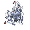
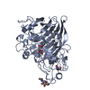
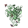
| Entry | Database: PDB / ID: 3ovw | |||||||||
|---|---|---|---|---|---|---|---|---|---|---|
| Title | ENDOGLUCANASE I NATIVE STRUCTURE | |||||||||
 Components Components | ENDOGLUCANASE I | |||||||||
 Keywords Keywords | HYDROLASE / GLYCOSYL HYDROLASE / ENDOGLUCANASE I / GLYCOSYLATED PROTEIN | |||||||||
| Function / homology |  Function and homology information Function and homology information | |||||||||
| Biological species |   Fusarium oxysporum (fungus) Fusarium oxysporum (fungus) | |||||||||
| Method |  X-RAY DIFFRACTION / MOLECULAR REPLACEMENT / Resolution: 2.3 Å X-RAY DIFFRACTION / MOLECULAR REPLACEMENT / Resolution: 2.3 Å | |||||||||
 Authors Authors | Davies, G.J. / Schulein, M. | |||||||||
 Citation Citation | Journal: Biochemistry / Year: 1997 Title: Structure of the endoglucanase I from Fusarium oxysporum: native, cellobiose, and 3,4-epoxybutyl beta-D-cellobioside-inhibited forms, at 2.3 A resolution. Authors: Sulzenbacher, G. / Schulein, M. / Davies, G.J. #1: Journal: Gene / Year: 1994Title: The Use of Conserved Cellulase Family-Specific Sequences to Clone Cellulase Homologue Cdnas from Fusarium Oxysporum Authors: Sheppard, P.O. / Grant, F.J. / Oort, P.J. / Sprecher, C.A. / Foster, D.C. / Hagen, F.S. / Upshall, A. / Mcknight, G.L. / O'Hara, P.J. | |||||||||
| History |
|
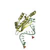
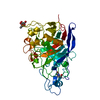
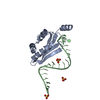
- Structure visualization
| Structure viewer | Molecule: MolmilJmol/JSmol |
|---|
- Downloads & links
-Download
| PDBx/mmCIF format | 3ovw.cif.gz | 172 KB | Display | PDBx/mmCIF format |
|---|---|---|---|---|
| PDB format | pdb3ovw.ent.gz | 135.6 KB | Display | PDB format |
| PDBx/mmJSON format | 3ovw.json.gz | Tree view | PDBx/mmJSON format | |
| Others |  Other downloads Other downloads |
-Validation report
| Arichive directory | https://data.pdbj.org/pub/pdb/validation_reports/ov/3ovwftp://data.pdbj.org/pub/pdb/validation_reports/ov/3ovw | HTTPS FTP |
|---|
-Related structure data


-Links
 PDBj
PDBj- Assembly
| Deposited unit | 
| ||||||||
|---|---|---|---|---|---|---|---|---|---|
| 1 | 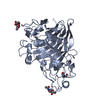
| ||||||||
| 2 | 
| ||||||||
| Unit cell |
|
-Components
| #1: Protein | Mass: 44685.320 Da / Num. of mol.: 2 / Source method: isolated from a natural source / Source: (natural) Fusarium oxysporum (fungus) / References: UniProt: P46237, cellulase#2: Sugar | ChemComp-NAG /   Type: D-saccharide, beta linking / Mass: 221.208 Da / Num. of mol.: 4 Type: D-saccharide, beta linking / Mass: 221.208 Da / Num. of mol.: 4Source method: isolated from a genetically manipulated source Formula: C8H15NO6 #3: Water | ChemComp-HOH / |  Mass: 18.015 Da / Num. of mol.: 422 / Source method: isolated from a natural source / Formula: H2O Mass: 18.015 Da / Num. of mol.: 422 / Source method: isolated from a natural source / Formula: H2OCompound details | THIS STRUCTURE BELONGS TO FAMILY 7 OF GLYCOSYL HYDROLASES. THIS IS THE NATIVE ENZYME STRUCTURE. THE ...THIS STRUCTURE BELONGS TO FAMILY 7 OF GLYCOSYL HYDROLASES | Has protein modification | Y | |
|---|
-Experimental details
-Experiment
| Experiment | Method: X-RAY DIFFRACTION / Number of used crystals: 1 |
|---|
- Sample preparation
| Crystal | Density Matthews: 2.37 Å3/Da / Density % sol: 48 % | ||||||||||||||||||||||||||||
|---|---|---|---|---|---|---|---|---|---|---|---|---|---|---|---|---|---|---|---|---|---|---|---|---|---|---|---|---|---|
| Crystal grow | Method: vapor diffusion, hanging drop / pH: 6.5 Details: 20 % PEG 8K, 0.2 M MAGNESIUM CHLORIDE, PH 6.5 FOR 0.1 M MOPS. METHOD: HANGING DROP VAPOR DIFFUSION, vapor diffusion - hanging drop | ||||||||||||||||||||||||||||
| Crystal grow | *PLUS Temperature: 16 ℃ / Method: vapor diffusion, hanging drop | ||||||||||||||||||||||||||||
| Components of the solutions | *PLUS
|
-Data collection
| Diffraction | Mean temperature: 293 K |
|---|---|
| Diffraction source | Source: ROTATING ANODE / Type: RIGAKU FR-C / Wavelength: 1.5418 |
| Detector | Date: 1994 |
| Radiation | Monochromatic (M) / Laue (L): M / Scattering type: x-ray |
| Radiation wavelength | Wavelength: 1.5418 Å / Relative weight: 1 |
| Reflection | Resolution: 2.3→15 Å / Num. obs: 32773 / % possible obs: 91 % / Redundancy: 2.4 % / Rmerge(I) obs: 0.072 |
| Reflection shell | Resolution: 2.3→2.4 Å / Redundancy: 1.87 % / Rmerge(I) obs: 0.185 / % possible all: 51.9 |
| Reflection shell | *PLUS % possible obs: 51 % |
- Processing
| Software |
| ||||||||||||||||||||||||||||||||||||||||||||||||||||||||||||||||||||||||||||||||||||
|---|---|---|---|---|---|---|---|---|---|---|---|---|---|---|---|---|---|---|---|---|---|---|---|---|---|---|---|---|---|---|---|---|---|---|---|---|---|---|---|---|---|---|---|---|---|---|---|---|---|---|---|---|---|---|---|---|---|---|---|---|---|---|---|---|---|---|---|---|---|---|---|---|---|---|---|---|---|---|---|---|---|---|---|---|---|
| Refinement | Method to determine structure: MOLECULAR REPLACEMENT Starting model: NATIVE STRUCTURE OF HUMICOLA INSOLENS EG I Resolution: 2.3→18 Å / σ(F): 0
| ||||||||||||||||||||||||||||||||||||||||||||||||||||||||||||||||||||||||||||||||||||
| Refinement step | Cycle: LAST / Resolution: 2.3→18 Å
| ||||||||||||||||||||||||||||||||||||||||||||||||||||||||||||||||||||||||||||||||||||
| Refine LS restraints |
| ||||||||||||||||||||||||||||||||||||||||||||||||||||||||||||||||||||||||||||||||||||
| Software | *PLUS Name: REFMAC / Classification: refinement | ||||||||||||||||||||||||||||||||||||||||||||||||||||||||||||||||||||||||||||||||||||
| Refinement | *PLUS Num. reflection all: 32757 / Rfactor all: 0.158 | ||||||||||||||||||||||||||||||||||||||||||||||||||||||||||||||||||||||||||||||||||||
| Solvent computation | *PLUS | ||||||||||||||||||||||||||||||||||||||||||||||||||||||||||||||||||||||||||||||||||||
| Displacement parameters | *PLUS |
