 Movie
Movie Controller

[English] 日本語
 Yorodumi
- PDB-3w15: Structure of peroxisomal targeting signal 2 (PTS2) of Saccharomyc... -
+ Open data

- Basic information
| Entry | Database: PDB / ID: 3w15 | ||||||
|---|---|---|---|---|---|---|---|
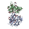
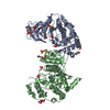
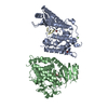
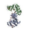
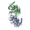
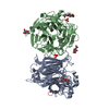
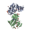
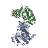
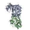
| Title | Structure of peroxisomal targeting signal 2 (PTS2) of Saccharomyces cerevisiae 3-ketoacyl-CoA thiolase in complex with Pex7p and Pex21p | ||||||
 Components Components |
| ||||||
 Keywords Keywords | PROTEIN TRANSPORT / beta-propeller / targeting signal recognition / cytosol / peroxisome | ||||||
| Function / homology |  Function and homology information Function and homology informationperoxisome matrix targeting signal-2 binding / cargo receptor complex / alpha-linolenic acid (ALA) metabolism / Beta-oxidation of very long chain fatty acids / acetyl-CoA C-acyltransferase / protein import into peroxisome matrix / Peroxisomal protein import / protein import into peroxisome matrix, docking / acetyl-CoA C-acyltransferase activity / phenylacetate catabolic process ...peroxisome matrix targeting signal-2 binding / cargo receptor complex / alpha-linolenic acid (ALA) metabolism / Beta-oxidation of very long chain fatty acids / acetyl-CoA C-acyltransferase / protein import into peroxisome matrix / Peroxisomal protein import / protein import into peroxisome matrix, docking / acetyl-CoA C-acyltransferase activity / phenylacetate catabolic process / protein carrier activity / peroxisomal membrane / fatty acid beta-oxidation / detection of maltose stimulus / maltose transport complex / peroxisomal matrix / carbohydrate transport / carbohydrate transmembrane transporter activity / maltose binding / maltose transport / maltodextrin transmembrane transport / ATP-binding cassette (ABC) transporter complex, substrate-binding subunit-containing / Neutrophil degranulation / ATP-binding cassette (ABC) transporter complex / cell chemotaxis / mitochondrial intermembrane space / peroxisome / outer membrane-bounded periplasmic space / periplasmic space / mRNA binding / DNA damage response / membrane / cytosol / cytoplasm Similarity search - Function | ||||||
| Biological species |   | ||||||
| Method |  X-RAY DIFFRACTION / SYNCHROTRON / MOLECULAR REPLACEMENT / molecular replacement / Resolution: 1.8 Å X-RAY DIFFRACTION / SYNCHROTRON / MOLECULAR REPLACEMENT / molecular replacement / Resolution: 1.8 Å | ||||||
 Authors Authors | Pan, D. / Nakatsu, T. / Kato, H. | ||||||
 Citation Citation | Journal: Nat.Struct.Mol.Biol. / Year: 2013 Title: Crystal structure of peroxisomal targeting signal-2 bound to its receptor complex Pex7p-Pex21p Authors: Pan, D. / Nakatsu, T. / Kato, H. | ||||||
| History |
|
- Structure visualization
| Structure viewer | Molecule: MolmilJmol/JSmol |
|---|
- Downloads & links
-Download
| PDBx/mmCIF format | 3w15.cif.gz | 187.7 KB | Display | PDBx/mmCIF format |
|---|---|---|---|---|
| PDB format | pdb3w15.ent.gz | 141.9 KB | Display | PDB format |
| PDBx/mmJSON format | 3w15.json.gz | Tree view | PDBx/mmJSON format | |
| Others |  Other downloads Other downloads |
-Validation report
| Arichive directory | https://data.pdbj.org/pub/pdb/validation_reports/w1/3w15ftp://data.pdbj.org/pub/pdb/validation_reports/w1/3w15 | HTTPS FTP |
|---|
-Related structure data
| Similar structure data |
|---|

-Links
 PDBj
PDBj


- Assembly
| Deposited unit | 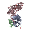
| ||||||||
|---|---|---|---|---|---|---|---|---|---|
| 1 |
| ||||||||
| Unit cell |
|
-Components
-Protein , 3 types, 3 molecules ABC
| #1: Protein | Mass: 41525.809 Da / Num. of mol.: 1 / Mutation: del(E257-V265) Source method: isolated from a genetically manipulated source Source: (gene. exp.) Strain: S288c / Gene: PEX7 / Plasmid: pPICZA / Production host:  Komagataella pastoris (fungus) / Strain (production host): SMD1163 / References: UniProt: P39108 Komagataella pastoris (fungus) / Strain (production host): SMD1163 / References: UniProt: P39108 |
|---|---|
| #2: Protein | Mass: 11004.136 Da / Num. of mol.: 1 / Fragment: UNP residue 190-288 Source method: isolated from a genetically manipulated source Source: (gene. exp.) Strain: S288c / Gene: PEX21 / Plasmid: pGEX-6P-1 / Production host: |
| #3: Protein | Mass: 42897.555 Da / Num. of mol.: 1 / Fragment: UNP residue 1-15, UNP residues 27-396 Source method: isolated from a genetically manipulated source Details: chimera of 3-ketoacyl-CoA thiolase, peroxisomal, Maltose-binding periplasmic protein Source: (gene. exp.) Strain: S288c, K12 / Gene: FOX3 / Plasmid: pGEX-6P-1 / Production host: |
-Non-polymers , 3 types, 578 molecules 


| #4: Chemical | ChemComp-NO3 /  Mass: 62.005 Da / Num. of mol.: 9 / Source method: obtained synthetically / Formula: NO3 Mass: 62.005 Da / Num. of mol.: 9 / Source method: obtained synthetically / Formula: NO3#5: Chemical | ChemComp-MG /  Mass: 24.305 Da / Num. of mol.: 6 / Source method: obtained synthetically / Formula: Mg Mass: 24.305 Da / Num. of mol.: 6 / Source method: obtained synthetically / Formula: Mg#6: Water | ChemComp-HOH / | Mass: 18.015 Da / Num. of mol.: 563 / Source method: isolated from a natural source / Formula: H2O |
|---|
-Details
| Has protein modification | Y |
|---|
-Experimental details
-Experiment
| Experiment | Method: X-RAY DIFFRACTION / Number of used crystals: 1 |
|---|
- Sample preparation
| Crystal | Density Matthews: 2.04 Å3/Da / Density % sol: 39.67 % / Mosaicity: 0.879 ° |
|---|---|
| Crystal grow | Temperature: 293 K / Method: vapor diffusion, hanging drop / pH: 7.7 Details: 25% PEG2000, 0.3M Magnesium nitrate, 0.1M Tris-HCl, pH 7.7, VAPOR DIFFUSION, HANGING DROP, temperature 293K |
-Data collection
| Diffraction | Mean temperature: 100 K | |||||||||||||||||||||||||||||||||||||||||||||||||||||||||||||||||||||||||||||
|---|---|---|---|---|---|---|---|---|---|---|---|---|---|---|---|---|---|---|---|---|---|---|---|---|---|---|---|---|---|---|---|---|---|---|---|---|---|---|---|---|---|---|---|---|---|---|---|---|---|---|---|---|---|---|---|---|---|---|---|---|---|---|---|---|---|---|---|---|---|---|---|---|---|---|---|---|---|---|
| Diffraction source | Source: SYNCHROTRON / Site: SPring-8  / Beamline: BL41XU / Wavelength: 1 Å / Beamline: BL41XU / Wavelength: 1 Å | |||||||||||||||||||||||||||||||||||||||||||||||||||||||||||||||||||||||||||||
| Detector | Type: MARMOSAIC 225 mm CCD / Detector: CCD / Date: Apr 15, 2011 | |||||||||||||||||||||||||||||||||||||||||||||||||||||||||||||||||||||||||||||
| Radiation | Protocol: SINGLE WAVELENGTH / Monochromatic (M) / Laue (L): M / Scattering type: x-ray | |||||||||||||||||||||||||||||||||||||||||||||||||||||||||||||||||||||||||||||
| Radiation wavelength | Wavelength: 1 Å / Relative weight: 1 | |||||||||||||||||||||||||||||||||||||||||||||||||||||||||||||||||||||||||||||
| Reflection | Resolution: 1.8→32.24 Å / Num. all: 71460 / Num. obs: 71460 / % possible obs: 99.77 % / Redundancy: 7.12 % / Rmerge(I) obs: 0.066 / Net I/σ(I): 20.2139 | |||||||||||||||||||||||||||||||||||||||||||||||||||||||||||||||||||||||||||||
| Reflection shell | Diffraction-ID: 1
|
-Phasing
| Phasing | Method: molecular replacement | |||||||||
|---|---|---|---|---|---|---|---|---|---|---|
| Phasing MR | Rfactor: 52.08 / Model details: Phaser MODE: MR_AUTO
|
- Processing
| Software |
| |||||||||||||||||||||||||||||||||||||||||||||||||||||||||||||||||||||||||||||||||||||||||||||||||||||||||||||||||||||||||||||||||||||||||||||||||||
|---|---|---|---|---|---|---|---|---|---|---|---|---|---|---|---|---|---|---|---|---|---|---|---|---|---|---|---|---|---|---|---|---|---|---|---|---|---|---|---|---|---|---|---|---|---|---|---|---|---|---|---|---|---|---|---|---|---|---|---|---|---|---|---|---|---|---|---|---|---|---|---|---|---|---|---|---|---|---|---|---|---|---|---|---|---|---|---|---|---|---|---|---|---|---|---|---|---|---|---|---|---|---|---|---|---|---|---|---|---|---|---|---|---|---|---|---|---|---|---|---|---|---|---|---|---|---|---|---|---|---|---|---|---|---|---|---|---|---|---|---|---|---|---|---|---|---|---|---|
| Refinement | Method to determine structure: MOLECULAR REPLACEMENT / Resolution: 1.8→32.2 Å / Cor.coef. Fo:Fc: 0.949 / Cor.coef. Fo:Fc free: 0.926 / WRfactor Rfree: 0.241 / WRfactor Rwork: 0.206 / Occupancy max: 1 / Occupancy min: 0.5 / SU B: 2.576 / SU ML: 0.083 / Cross valid method: THROUGHOUT / σ(F): 0 / ESU R: 0.142 / ESU R Free: 0.13 / Stereochemistry target values: MAXIMUM LIKELIHOOD Details: HYDROGENS HAVE BEEN ADDED IN THE RIDING POSITIONS U VALUES: REFINED INDIVIDUALLY
| |||||||||||||||||||||||||||||||||||||||||||||||||||||||||||||||||||||||||||||||||||||||||||||||||||||||||||||||||||||||||||||||||||||||||||||||||||
| Solvent computation | Ion probe radii: 0.8 Å / Shrinkage radii: 0.8 Å / VDW probe radii: 1.4 Å / Solvent model: MASK | |||||||||||||||||||||||||||||||||||||||||||||||||||||||||||||||||||||||||||||||||||||||||||||||||||||||||||||||||||||||||||||||||||||||||||||||||||
| Displacement parameters | Biso max: 52.01 Å2 / Biso mean: 21.4142 Å2 / Biso min: 7.9 Å2
| |||||||||||||||||||||||||||||||||||||||||||||||||||||||||||||||||||||||||||||||||||||||||||||||||||||||||||||||||||||||||||||||||||||||||||||||||||
| Refinement step | Cycle: LAST / Resolution: 1.8→32.2 Å
| |||||||||||||||||||||||||||||||||||||||||||||||||||||||||||||||||||||||||||||||||||||||||||||||||||||||||||||||||||||||||||||||||||||||||||||||||||
| Refine LS restraints |
| |||||||||||||||||||||||||||||||||||||||||||||||||||||||||||||||||||||||||||||||||||||||||||||||||||||||||||||||||||||||||||||||||||||||||||||||||||
| LS refinement shell | Refine-ID: X-RAY DIFFRACTION / Total num. of bins used: 20
|
