 Movie
Movie Controller

[English] 日本語
 Yorodumi
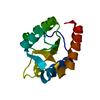
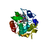
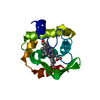
- PDB-3vnw: Crystal structure of cytochrome c552 from Thermus thermophilus at... -
+ Open data

- Basic information
| Entry | Database: PDB / ID: 3vnw | ||||||
|---|---|---|---|---|---|---|---|
| Title | Crystal structure of cytochrome c552 from Thermus thermophilus at pH 5.44 | ||||||
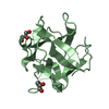
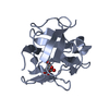
 Components Components | Cytochrome c-552 | ||||||
 Keywords Keywords | ELECTRON TRANSPORT / Cytochrome c / Electron Transfer | ||||||
| Function / homology |  Function and homology information Function and homology information | ||||||
| Biological species |   Thermus thermophilus (bacteria) Thermus thermophilus (bacteria) | ||||||
| Method |  X-RAY DIFFRACTION / MOLECULAR REPLACEMENT / Resolution: 1.97 Å X-RAY DIFFRACTION / MOLECULAR REPLACEMENT / Resolution: 1.97 Å | ||||||
 Authors Authors | Yamada, S. / Bouley-Ford, N.D. / Keller, G.E. / Winkler, J.R. / Gray, H.B. | ||||||
 Citation Citation | Journal: Proc.Natl.Acad.Sci.USA / Year: 2013 Title: Snapshots of a protein folding intermediate Authors: Yamada, S. / Bouley-Ford, N.D. / Keller, G.E. / Ford, W.C. / Gray, H.B. / Winkler, J.R. | ||||||
| History |
|
- Structure visualization
| Structure viewer | Molecule: MolmilJmol/JSmol |
|---|
- Downloads & links
-Download
| PDBx/mmCIF format | 3vnw.cif.gz | 40.8 KB | Display | PDBx/mmCIF format |
|---|---|---|---|---|
| PDB format | pdb3vnw.ent.gz | 27.2 KB | Display | PDB format |
| PDBx/mmJSON format | 3vnw.json.gz | Tree view | PDBx/mmJSON format | |
| Others |  Other downloads Other downloads |
-Validation report
| Arichive directory | https://data.pdbj.org/pub/pdb/validation_reports/vn/3vnwftp://data.pdbj.org/pub/pdb/validation_reports/vn/3vnw | HTTPS FTP |
|---|
-Related structure data
| Related structure data | 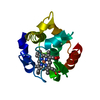 1dt1S S: Starting model for refinement |
|---|---|
| Similar structure data |


-Links
 PDBj
PDBj


- Assembly
| Deposited unit | 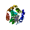
| ||||||||
|---|---|---|---|---|---|---|---|---|---|
| 1 |
| ||||||||
| Unit cell |
|
-Components
| #1: Protein | Mass: 13995.368 Da / Num. of mol.: 1 Source method: isolated from a genetically manipulated source Source: (gene. exp.) Thermus thermophilus (bacteria) / Gene: cycA / Plasmid: pET17b / Production host: |
|---|---|
| #2: Chemical | ChemComp-HEM /   Mass: 616.487 Da / Num. of mol.: 1 / Source method: obtained synthetically / Formula: C34H32FeN4O4 Mass: 616.487 Da / Num. of mol.: 1 / Source method: obtained synthetically / Formula: C34H32FeN4O4 |
| #3: Water | ChemComp-HOH /  Mass: 18.015 Da / Num. of mol.: 71 / Source method: isolated from a natural source / Formula: H2O Mass: 18.015 Da / Num. of mol.: 71 / Source method: isolated from a natural source / Formula: H2O |
-Experimental details
-Experiment
| Experiment | Method: X-RAY DIFFRACTION / Number of used crystals: 1 |
|---|
- Sample preparation
| Crystal | Density Matthews: 2.45 Å3/Da / Density % sol: 49.75 % |
|---|---|
| Crystal grow | Temperature: 298 K / Method: vapor diffusion, sitting drop / pH: 5.44 Details: 0.2M Imidazole-Malate, 42% MPEG 5K, pH 5.44, VAPOR DIFFUSION, SITTING DROP, temperature 298K |
-Data collection
| Diffraction | Mean temperature: 100 K |
|---|---|
| Diffraction source | Source: ROTATING ANODE / Type: RIGAKU MICROMAX-007 HF / Wavelength: 1.5418 Å |
| Detector | Type: RIGAKU RAXIS IV / Detector: IMAGE PLATE / Date: Jul 15, 2011 |
| Radiation | Protocol: SINGLE WAVELENGTH / Monochromatic (M) / Laue (L): M / Scattering type: x-ray |
| Radiation wavelength | Wavelength: 1.5418 Å / Relative weight: 1 |
| Reflection | Resolution: 1.97→25.68 Å / Num. all: 9951 / Num. obs: 9814 / % possible obs: 98.6 % / Observed criterion σ(F): 2 / Observed criterion σ(I): 2 / Redundancy: 4.8 % / Rmerge(I) obs: 0.088 / Net I/σ(I): 9.7 |
| Reflection shell | Resolution: 1.97→2.04 Å / Redundancy: 4.8 % / Rmerge(I) obs: 0.312 / Mean I/σ(I) obs: 3.6 / Num. unique all: 956 / % possible all: 98.4 |
- Processing
| Software |
| |||||||||||||||||||||||||||||||||||||||||||||||||||||||||||||||||
|---|---|---|---|---|---|---|---|---|---|---|---|---|---|---|---|---|---|---|---|---|---|---|---|---|---|---|---|---|---|---|---|---|---|---|---|---|---|---|---|---|---|---|---|---|---|---|---|---|---|---|---|---|---|---|---|---|---|---|---|---|---|---|---|---|---|---|
| Refinement | Method to determine structure: MOLECULAR REPLACEMENT Starting model: PDB ENTRY 1DT1 Resolution: 1.97→20 Å / Cor.coef. Fo:Fc: 0.939 / Cor.coef. Fo:Fc free: 0.934 / SU B: 3.903 / SU ML: 0.114 / Cross valid method: THROUGHOUT / ESU R: 0.202 / ESU R Free: 0.16 / Stereochemistry target values: MAXIMUM LIKELIHOOD
| |||||||||||||||||||||||||||||||||||||||||||||||||||||||||||||||||
| Solvent computation | Ion probe radii: 0.8 Å / Shrinkage radii: 0.8 Å / VDW probe radii: 1.4 Å / Solvent model: MASK | |||||||||||||||||||||||||||||||||||||||||||||||||||||||||||||||||
| Displacement parameters | Biso mean: 22.245 Å2 | |||||||||||||||||||||||||||||||||||||||||||||||||||||||||||||||||
| Refinement step | Cycle: LAST / Resolution: 1.97→20 Å
| |||||||||||||||||||||||||||||||||||||||||||||||||||||||||||||||||
| Refine LS restraints |
| |||||||||||||||||||||||||||||||||||||||||||||||||||||||||||||||||
| LS refinement shell | Resolution: 1.97→2.021 Å / Total num. of bins used: 20
|
