 Movie
Movie Controller

[English] 日本語
 Yorodumi
- PDB-1c52: THERMUS THERMOPHILUS CYTOCHROME-C552: A NEW HIGHLY THERMOSTABLE C... -
+ Open data

- Basic information
| Entry | Database: PDB / ID: 1c52 | ||||||
|---|---|---|---|---|---|---|---|
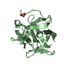
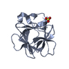
| Title | THERMUS THERMOPHILUS CYTOCHROME-C552: A NEW HIGHLY THERMOSTABLE CYTOCHROME-C STRUCTURE OBTAINED BY MAD PHASING | ||||||
 Components Components | CYTOCHROME-C552 | ||||||
 Keywords Keywords | ELECTRON TRANSPORT PROTEIN / CYTOCHROME-C552 / MAD / THERMOSTABILITY | ||||||
| Function / homology |  Function and homology information Function and homology information | ||||||
| Biological species |   Thermus thermophilus (bacteria) Thermus thermophilus (bacteria) | ||||||
| Method |  X-RAY DIFFRACTION / SYNCHROTRON / MAD / Resolution: 1.28 Å X-RAY DIFFRACTION / SYNCHROTRON / MAD / Resolution: 1.28 Å | ||||||
 Authors Authors | Than, M.E. / Hof, P. / Huber, R. / Bourenkov, G.P. / Bartunik, H.D. / Buse, G. / Soulimane, T. | ||||||
 Citation Citation | Journal: J.Mol.Biol. / Year: 1997 Title: Thermus thermophilus cytochrome-c552: A new highly thermostable cytochrome-c structure obtained by MAD phasing. Authors: Than, M.E. / Hof, P. / Huber, R. / Bourenkov, G.P. / Bartunik, H.D. / Buse, G. / Soulimane, T. #1: Journal: Biochem.Biophys.Res.Commun. / Year: 1985Title: Amino Acid Sequence of Cytochrome C-552 from Thermus Thermophilus Hb8 Authors: Titani, K. / Ericsson, L.H. / Hon-Nami, K. / Miyazawa, T. #2: Journal: J.Mol.Biol. / Year: 1978Title: Reversible Thermal Unfolding of Thermostable Cytochrome C-552 Authors: Nojima, H. / Hon-Nami, K. / Oshima, T. / Noda, H. #3: Journal: J.Biochem.(Tokyo) / Year: 1977Title: Purification and Some Properties of Cytochrome C-552 from an Extreme Thermophile, Thermus Thermophilus Hb8 Authors: Hon-Nami, K. / Oshima, T. | ||||||
| History |
|
- Structure visualization
| Structure viewer | Molecule: MolmilJmol/JSmol |
|---|
- Downloads & links
-Download
| PDBx/mmCIF format | 1c52.cif.gz | 55.8 KB | Display | PDBx/mmCIF format |
|---|---|---|---|---|
| PDB format | pdb1c52.ent.gz | 40.4 KB | Display | PDB format |
| PDBx/mmJSON format | 1c52.json.gz | Tree view | PDBx/mmJSON format | |
| Others |  Other downloads Other downloads |
-Validation report
| Arichive directory | https://data.pdbj.org/pub/pdb/validation_reports/c5/1c52ftp://data.pdbj.org/pub/pdb/validation_reports/c5/1c52 | HTTPS FTP |
|---|
-Related structure data
| Similar structure data |
|---|


-Links
 PDBj
PDBj


- Assembly
| Deposited unit | 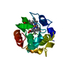
| ||||||||
|---|---|---|---|---|---|---|---|---|---|
| 1 |
| ||||||||
| Unit cell |
|
-Components
| #1: Protein | Mass: 14194.575 Da / Num. of mol.: 1 / Source method: isolated from a natural source / Details: OXIDIZED STATE (FE+++) / Source: (natural) Thermus thermophilus (bacteria) / Cellular location: PERIPLASM / References: UniProt: P04164 |
|---|---|
| #2: Chemical | ChemComp-HEM /   Mass: 616.487 Da / Num. of mol.: 1 / Source method: obtained synthetically / Formula: C34H32FeN4O4 Mass: 616.487 Da / Num. of mol.: 1 / Source method: obtained synthetically / Formula: C34H32FeN4O4 |
| #3: Water | ChemComp-HOH /  Mass: 18.015 Da / Num. of mol.: 196 / Source method: isolated from a natural source / Formula: H2O Mass: 18.015 Da / Num. of mol.: 196 / Source method: isolated from a natural source / Formula: H2O |
| Has protein modification | Y |
-Experimental details
-Experiment
| Experiment | Method: X-RAY DIFFRACTION / Number of used crystals: 1 |
|---|
- Sample preparation
| Crystal | Density Matthews: 2.58 Å3/Da / Density % sol: 52.3 % | |||||||||||||||||||||||||
|---|---|---|---|---|---|---|---|---|---|---|---|---|---|---|---|---|---|---|---|---|---|---|---|---|---|---|
| Crystal grow | pH: 8.2 Details: PROTEIN WAS CRYSTALLIZED FROM 2.8 M AMMONIUM SULFATE, 100 MM TRIS/HCL, PH 8.2 | |||||||||||||||||||||||||
| Crystal grow | *PLUS Method: unknownDetails: drop consists of 8 microlitter protein and 2 microlitter reservoir solution | |||||||||||||||||||||||||
| Components of the solutions | *PLUS
|
-Data collection
| Diffraction | Mean temperature: 90 K |
|---|---|
| Diffraction source | Source: SYNCHROTRON / Site: MPG/DESY, HAMBURG  / Beamline: BW6 / Wavelength: 1.05 / Beamline: BW6 / Wavelength: 1.05 |
| Detector | Type: MARRESEARCH / Detector: IMAGE PLATE / Date: Oct 1, 1996 / Details: MIRROR |
| Radiation | Monochromator: DOUBLE CRYSTAL SI(111) / Monochromatic (M) / Laue (L): M / Scattering type: x-ray |
| Radiation wavelength | Wavelength: 1.05 Å / Relative weight: 1 |
| Reflection | Resolution: 1.28→14.6 Å / Num. obs: 36004 / % possible obs: 93.6 % / Redundancy: 4.6 % / Biso Wilson estimate: 12.7 Å2 / Rsym value: 0.033 / Net I/σ(I): 14.1 |
| Reflection shell | Resolution: 1.28→1.31 Å / Redundancy: 2.5 % / Mean I/σ(I) obs: 1.6 / Rsym value: 0.357 / % possible all: 78.2 |
| Reflection | *PLUS Num. measured all: 165576 / Rmerge(I) obs: 0.033 |
| Reflection shell | *PLUS % possible obs: 78.2 % / Rmerge(I) obs: 0.357 |
- Processing
| Software |
| ||||||||||||||||||||||||||||||||||||||||||||||||||||||||||||
|---|---|---|---|---|---|---|---|---|---|---|---|---|---|---|---|---|---|---|---|---|---|---|---|---|---|---|---|---|---|---|---|---|---|---|---|---|---|---|---|---|---|---|---|---|---|---|---|---|---|---|---|---|---|---|---|---|---|---|---|---|---|
| Refinement | Method to determine structure: MAD / Resolution: 1.28→8 Å / Data cutoff high absF: 100000 / Data cutoff low absF: 0.1 / Isotropic thermal model: RESTRAINED Details: OCCUPANCY AND B-FACTOR ARE SET TO ZERO FOR ALL ATOMS THAT ARE NOT DEFINED IN THE FINAL 2FO-FC ELECTRON DENSITY.
| ||||||||||||||||||||||||||||||||||||||||||||||||||||||||||||
| Displacement parameters | Biso mean: 15.1 Å2 | ||||||||||||||||||||||||||||||||||||||||||||||||||||||||||||
| Refinement step | Cycle: LAST / Resolution: 1.28→8 Å
| ||||||||||||||||||||||||||||||||||||||||||||||||||||||||||||
| Refine LS restraints |
| ||||||||||||||||||||||||||||||||||||||||||||||||||||||||||||
| LS refinement shell | Resolution: 1.28→1.29 Å / Total num. of bins used: 30
| ||||||||||||||||||||||||||||||||||||||||||||||||||||||||||||
| Software | *PLUS Name: X-PLOR / Version: 3.851 / Classification: refinement | ||||||||||||||||||||||||||||||||||||||||||||||||||||||||||||
| Refinement | *PLUS | ||||||||||||||||||||||||||||||||||||||||||||||||||||||||||||
| Solvent computation | *PLUS | ||||||||||||||||||||||||||||||||||||||||||||||||||||||||||||
| Displacement parameters | *PLUS | ||||||||||||||||||||||||||||||||||||||||||||||||||||||||||||
| Refine LS restraints | *PLUS
| ||||||||||||||||||||||||||||||||||||||||||||||||||||||||||||
| LS refinement shell | *PLUS Rfactor obs: 0.327 |
