 Movie
Movie Controller

[English] 日本語
 Yorodumi
- PDB-3vmt: Crystal structure of Staphylococcus aureus membrane-bound transgl... -
+ Open data

- Basic information
| Entry | Database: PDB / ID: 3vmt | ||||||
|---|---|---|---|---|---|---|---|
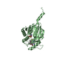
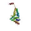
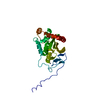
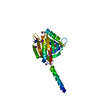
| Title | Crystal structure of Staphylococcus aureus membrane-bound transglycosylase in complex with a Lipid II analog | ||||||
 Components Components | Monofunctional glycosyltransferase | ||||||
 Keywords Keywords | TRANSFERASE / Transmembrane / Glycosyltransferase / Bacterial cell wall synthesis / Membrane | ||||||
| Function / homology |  Function and homology information Function and homology informationpeptidoglycan glycosyltransferase / peptidoglycan glycosyltransferase activity / peptidoglycan biosynthetic process / cell wall organization / regulation of cell shape / outer membrane-bounded periplasmic space / plasma membrane Similarity search - Function | ||||||
| Biological species |   Staphylococcus aureus (bacteria) Staphylococcus aureus (bacteria) | ||||||
| Method |  X-RAY DIFFRACTION / SYNCHROTRON / MOLECULAR REPLACEMENT / Resolution: 2.299 Å X-RAY DIFFRACTION / SYNCHROTRON / MOLECULAR REPLACEMENT / Resolution: 2.299 Å | ||||||
 Authors Authors | Huang, C.Y. / Shih, H.W. / Lin, L.Y. / Tien, Y.W. / Cheng, T.J.R. / Cheng, W.C. / Wong, C.H. / Ma, C. | ||||||
 Citation Citation | Journal: Proc.Natl.Acad.Sci.USA / Year: 2012 Title: Crystal structure of Staphylococcus aureus transglycosylase in complex with a lipid II analog and elucidation of peptidoglycan synthesis mechanism Authors: Huang, C.Y. / Shih, H.W. / Lin, L.Y. / Tien, Y.W. / Cheng, T.J.R. / Cheng, W.C. / Wong, C.H. / Ma, C. | ||||||
| History |
|
- Structure visualization
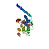
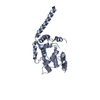
| Structure viewer | Molecule: MolmilJmol/JSmol |
|---|
- Downloads & links
-Download
| PDBx/mmCIF format | 3vmt.cif.gz | 200.7 KB | Display | PDBx/mmCIF format |
|---|---|---|---|---|
| PDB format | pdb3vmt.ent.gz | 157.7 KB | Display | PDB format |
| PDBx/mmJSON format | 3vmt.json.gz | Tree view | PDBx/mmJSON format | |
| Others |  Other downloads Other downloads |
-Validation report
| Arichive directory | https://data.pdbj.org/pub/pdb/validation_reports/vm/3vmtftp://data.pdbj.org/pub/pdb/validation_reports/vm/3vmt | HTTPS FTP |
|---|
-Related structure data
| Related structure data |  3vmqC  3vmrC  3vmsC 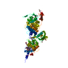 3fwlS  3fwm 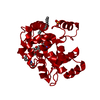 3hzsS C: citing same article ( S: Starting model for refinement |
|---|---|
| Similar structure data |


-Links
 PDBj
PDBj- Assembly
| Deposited unit | 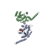
| ||||||||
|---|---|---|---|---|---|---|---|---|---|
| 1 | 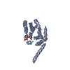
| ||||||||
| 2 | 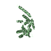
| ||||||||
| Unit cell |
|
-Components
| #1: Protein | Mass: 30314.369 Da / Num. of mol.: 2 / Fragment: residues 28-269 Source method: isolated from a genetically manipulated source Source: (gene. exp.) Staphylococcus aureus (bacteria) / Strain: Mu50 / Gene: MGT / Plasmid: pET15b / Production host: References: UniProt: Q99T05, Transferases; Glycosyltransferases #2: Chemical | ChemComp-LHI / [( | 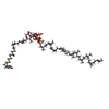  Mass: 1833.271 Da / Num. of mol.: 1 / Source method: obtained synthetically / Formula: C93H155N7O23P2S Mass: 1833.271 Da / Num. of mol.: 1 / Source method: obtained synthetically / Formula: C93H155N7O23P2S#3: Chemical |   Mass: 24.305 Da / Num. of mol.: 2 / Source method: obtained synthetically / Formula: Mg Mass: 24.305 Da / Num. of mol.: 2 / Source method: obtained synthetically / Formula: Mg#4: Water | ChemComp-HOH / |  Mass: 18.015 Da / Num. of mol.: 52 / Source method: isolated from a natural source / Formula: H2O Mass: 18.015 Da / Num. of mol.: 52 / Source method: isolated from a natural source / Formula: H2ONonpolymer details | THE LIGAND LHI IS THE ANALOG OF LIPIDII(LIIA). | |
|---|
-Experimental details
-Experiment
| Experiment | Method: X-RAY DIFFRACTION / Number of used crystals: 1 |
|---|
- Sample preparation
| Crystal | Density Matthews: 2.81 Å3/Da / Density % sol: 56.28 % |
|---|---|
| Crystal grow | Temperature: 277 K / Method: vapor diffusion, hanging drop / pH: 8 Details: 100mM MgCl2, 100mM HEPES, 25% PEG400, pH 8.0, VAPOR DIFFUSION, HANGING DROP, temperature 277K |
-Data collection
| Diffraction | Mean temperature: 100 K |
|---|---|
| Diffraction source | Source: SYNCHROTRON / Site: SPring-8  / Beamline: BL44XU / Wavelength: 1 Å / Beamline: BL44XU / Wavelength: 1 Å |
| Detector | Type: Bruker DIP-6040 / Detector: CCD / Date: Oct 20, 2011 |
| Radiation | Protocol: SINGLE WAVELENGTH / Monochromatic (M) / Laue (L): M / Scattering type: x-ray |
| Radiation wavelength | Wavelength: 1 Å / Relative weight: 1 |
| Reflection | Resolution: 2.29→29.26 Å / Num. obs: 30750 / % possible obs: 98 % / Observed criterion σ(F): 2 / Observed criterion σ(I): 1 / Biso Wilson estimate: 43.19 Å2 |
| Reflection shell | Resolution: 2.2987→2.3808 Å |
- Processing
| Software |
| |||||||||||||||||||||||||||||||||||||||||||||||||||||||||||||||||||||||||||||
|---|---|---|---|---|---|---|---|---|---|---|---|---|---|---|---|---|---|---|---|---|---|---|---|---|---|---|---|---|---|---|---|---|---|---|---|---|---|---|---|---|---|---|---|---|---|---|---|---|---|---|---|---|---|---|---|---|---|---|---|---|---|---|---|---|---|---|---|---|---|---|---|---|---|---|---|---|---|---|
| Refinement | Method to determine structure: MOLECULAR REPLACEMENT Starting model: 3HZS, 3FWM, 3FWL Resolution: 2.299→28.079 Å / Occupancy max: 1 / Occupancy min: 1 / FOM work R set: 0.7624 / SU ML: 0.27 / σ(F): 0.02 / Phase error: 29.09 / Stereochemistry target values: ML
| |||||||||||||||||||||||||||||||||||||||||||||||||||||||||||||||||||||||||||||
| Solvent computation | Shrinkage radii: 0.9 Å / VDW probe radii: 1.11 Å / Solvent model: FLAT BULK SOLVENT MODEL / Bsol: 73.778 Å2 / ksol: 0.363 e/Å3 | |||||||||||||||||||||||||||||||||||||||||||||||||||||||||||||||||||||||||||||
| Displacement parameters | Biso max: 265.53 Å2 / Biso mean: 87.1801 Å2 / Biso min: 29.31 Å2
| |||||||||||||||||||||||||||||||||||||||||||||||||||||||||||||||||||||||||||||
| Refinement step | Cycle: LAST / Resolution: 2.299→28.079 Å
| |||||||||||||||||||||||||||||||||||||||||||||||||||||||||||||||||||||||||||||
| Refine LS restraints |
| |||||||||||||||||||||||||||||||||||||||||||||||||||||||||||||||||||||||||||||
| LS refinement shell | Refine-ID: X-RAY DIFFRACTION / Total num. of bins used: 10
| |||||||||||||||||||||||||||||||||||||||||||||||||||||||||||||||||||||||||||||
| Refinement TLS params. | Method: refined / Origin x: -24.2765 Å / Origin y: -1.1594 Å / Origin z: 4.8341 Å
| |||||||||||||||||||||||||||||||||||||||||||||||||||||||||||||||||||||||||||||
| Refinement TLS group |
|
