 Movie
Movie Controller

[English] 日本語
 Yorodumi
- PDB-3vjo: Crystal structure of the wild-type EGFR kinase domain in complex ... -
+ Open data

- Basic information
| Entry | Database: PDB / ID: 3vjo | ||||||
|---|---|---|---|---|---|---|---|
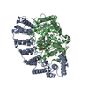
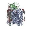
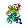
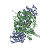
| Title | Crystal structure of the wild-type EGFR kinase domain in complex with AMPPNP. | ||||||
 Components Components | Epidermal growth factor receptor | ||||||
 Keywords Keywords | TRANSFERASE / receptor / disease mutation / cell cycle / drug resistance / kinase / tyrosine-protein kinase / ATP binding / phosphorylation / transmembrane | ||||||
| Function / homology |  Function and homology information Function and homology informationmultivesicular body, internal vesicle lumen / negative regulation of cardiocyte differentiation / Shc-EGFR complex / positive regulation of protein kinase C signaling / Inhibition of Signaling by Overexpressed EGFR / epidermal growth factor receptor activity / EGFR interacts with phospholipase C-gamma / epidermal growth factor binding / regulation of peptidyl-tyrosine phosphorylation / response to UV-A ...multivesicular body, internal vesicle lumen / negative regulation of cardiocyte differentiation / Shc-EGFR complex / positive regulation of protein kinase C signaling / Inhibition of Signaling by Overexpressed EGFR / epidermal growth factor receptor activity / EGFR interacts with phospholipase C-gamma / epidermal growth factor binding / regulation of peptidyl-tyrosine phosphorylation / response to UV-A / ubiquitin-dependent endocytosis / PLCG1 events in ERBB2 signaling / morphogenesis of an epithelial fold / PTK6 promotes HIF1A stabilization / ERBB2 Activates PTK6 Signaling / digestive tract morphogenesis / ERBB2-EGFR signaling pathway / Signaling by EGFR / eyelid development in camera-type eye / intracellular vesicle / cerebral cortex cell migration / ERBB2 Regulates Cell Motility / Developmental Lineage of Mammary Gland Myoepithelial Cells / protein insertion into membrane / protein tyrosine kinase activator activity / Signaling by ERBB4 / Respiratory syncytial virus (RSV) attachment and entry / negative regulation of epidermal growth factor receptor signaling pathway / PI3K events in ERBB2 signaling / hair follicle development / positive regulation of phosphorylation / positive regulation of peptidyl-serine phosphorylation / Estrogen-dependent nuclear events downstream of ESR-membrane signaling / MAP kinase kinase kinase activity / embryonic placenta development / GAB1 signalosome / salivary gland morphogenesis / xenobiotic transport / positive regulation of G1/S transition of mitotic cell cycle / positive regulation of epidermal growth factor receptor signaling pathway / Signaling by ERBB2 / TFAP2 (AP-2) family regulates transcription of growth factors and their receptors / transmembrane receptor protein tyrosine kinase activity / GRB2 events in EGFR signaling / SHC1 events in EGFR signaling / EGFR Transactivation by Gastrin / epithelial cell proliferation / GRB2 events in ERBB2 signaling / SHC1 events in ERBB2 signaling / basal plasma membrane / ossification / cellular response to epidermal growth factor stimulus / positive regulation of DNA replication / positive regulation of epithelial cell proliferation / positive regulation of DNA repair / Signal transduction by L1 / positive regulation of protein localization to plasma membrane / cellular response to estradiol stimulus / cellular response to amino acid stimulus / phosphatidylinositol 3-kinase/protein kinase B signal transduction / sperm end piece / NOTCH3 Activation and Transmission of Signal to the Nucleus / clathrin-coated endocytic vesicle membrane / Signaling by ERBB2 TMD/JMD mutants / EGFR downregulation / receptor protein-tyrosine kinase / Constitutive Signaling by EGFRvIII / cell-cell adhesion / negative regulation of protein catabolic process / Signaling by ERBB2 ECD mutants / Signaling by ERBB2 KD Mutants / positive regulation of protein phosphorylation / positive regulation of miRNA transcription / positive regulation of fibroblast proliferation / cell morphogenesis / epidermal growth factor receptor signaling pathway / ruffle membrane / kinase binding / Downregulation of ERBB2 signaling / Constitutive Signaling by Aberrant PI3K in Cancer / neuron differentiation / HCMV Early Events / actin filament binding / cell junction / transmembrane signaling receptor activity / PIP3 activates AKT signaling / positive regulation of canonical Wnt signaling pathway / Cargo recognition for clathrin-mediated endocytosis / Constitutive Signaling by Ligand-Responsive EGFR Cancer Variants / Clathrin-mediated endocytosis / ATPase binding / PI5P, PP2A and IER3 Regulate PI3K/AKT Signaling / sperm principal piece / virus receptor activity / positive regulation of cell growth / RAF/MAP kinase cascade / protein tyrosine kinase activity / double-stranded DNA binding / sperm midpiece / early endosome membrane Similarity search - Function | ||||||
| Biological species |  Homo sapiens (human) Homo sapiens (human) | ||||||
| Method |  X-RAY DIFFRACTION / SYNCHROTRON / MOLECULAR REPLACEMENT / Resolution: 2.64 Å X-RAY DIFFRACTION / SYNCHROTRON / MOLECULAR REPLACEMENT / Resolution: 2.64 Å | ||||||
 Authors Authors | Yoshikawa, S. / Kukimoto-Niino, M. / Shirouzu, M. / Semba, K. / Yamamoto, T. / Yokoyama, S. | ||||||
 Citation Citation | Journal: Oncogene / Year: 2013 Title: Structural basis for the altered drug sensitivities of non-small cell lung cancer-associated mutants of human epidermal growth factor receptor. Authors: Yoshikawa, S. / Kukimoto-Niino, M. / Parker, L. / Handa, N. / Terada, T. / Fujimoto, T. / Terazawa, Y. / Wakiyama, M. / Sato, M. / Sano, S. / Kobayashi, T. / Tanaka, T. / Chen, L. / Liu, Z.J. ...Authors: Yoshikawa, S. / Kukimoto-Niino, M. / Parker, L. / Handa, N. / Terada, T. / Fujimoto, T. / Terazawa, Y. / Wakiyama, M. / Sato, M. / Sano, S. / Kobayashi, T. / Tanaka, T. / Chen, L. / Liu, Z.J. / Wang, B.C. / Shirouzu, M. / Kawa, S. / Semba, K. / Yamamoto, T. / Yokoyama, S. | ||||||
| History |
|
- Structure visualization
| Structure viewer | Molecule: MolmilJmol/JSmol |
|---|
- Downloads & links
-Download
| PDBx/mmCIF format | 3vjo.cif.gz | 76.1 KB | Display | PDBx/mmCIF format |
|---|---|---|---|---|
| PDB format | pdb3vjo.ent.gz | 55.5 KB | Display | PDB format |
| PDBx/mmJSON format | 3vjo.json.gz | Tree view | PDBx/mmJSON format | |
| Others |  Other downloads Other downloads |
-Validation report
| Arichive directory | https://data.pdbj.org/pub/pdb/validation_reports/vj/3vjoftp://data.pdbj.org/pub/pdb/validation_reports/vj/3vjo | HTTPS FTP |
|---|
-Related structure data
| Related structure data | 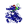 2eb2C  2eb3C 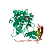 3ug1C 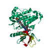 3ug2C  3vjnC 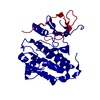 2gs2S C: citing same article ( S: Starting model for refinement |
|---|---|
| Similar structure data |
-Links
 PDBj
PDBj


- Assembly
| Deposited unit | 
| ||||||||
|---|---|---|---|---|---|---|---|---|---|
| 1 | 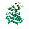
| ||||||||
| Unit cell |
|
-Components
| #1: Protein | Mass: 37977.930 Da / Num. of mol.: 1 / Fragment: kinase domain (UNP RESIDUES 695-1022) Source method: isolated from a genetically manipulated source Source: (gene. exp.) Homo sapiens (human) / Gene: EGFR, ERBB1 / Plasmid: pFastBac_HT_C / Cell line (production host): Sf9 / Production host:   Spodoptera frugiperda (fall armyworm) Spodoptera frugiperda (fall armyworm)References: UniProt: P00533, receptor protein-tyrosine kinase |
|---|---|
| #2: Chemical | ChemComp-ANP /   Mass: 506.196 Da / Num. of mol.: 1 / Source method: obtained synthetically / Formula: C10H17N6O12P3 / Comment: AMP-PNP, energy-carrying molecule analogue*YM Mass: 506.196 Da / Num. of mol.: 1 / Source method: obtained synthetically / Formula: C10H17N6O12P3 / Comment: AMP-PNP, energy-carrying molecule analogue*YM |
| #3: Water | ChemComp-HOH /  Mass: 18.015 Da / Num. of mol.: 9 / Source method: isolated from a natural source / Formula: H2O Mass: 18.015 Da / Num. of mol.: 9 / Source method: isolated from a natural source / Formula: H2O |
-Experimental details
-Experiment
| Experiment | Method: X-RAY DIFFRACTION / Number of used crystals: 1 |
|---|
- Sample preparation
| Crystal | Density Matthews: 3.23 Å3/Da / Density % sol: 61.95 % |
|---|---|
| Crystal grow | Temperature: 293 K / Method: vapor diffusion, sitting drop / pH: 7 Details: 0.1M Tris-HCl(pH 7.0), 0.2M NaCl, 1M Sodium citrate, VAPOR DIFFUSION, SITTING DROP, temperature 293K |
-Data collection
| Diffraction | Mean temperature: 100 K |
|---|---|
| Diffraction source | Source: SYNCHROTRON / Site: SLS  / Beamline: X06SA / Wavelength: 0.9786 Å / Beamline: X06SA / Wavelength: 0.9786 Å |
| Detector | Type: DECTRIS PILATUS 6M / Detector: PIXEL / Date: Aug 13, 2007 |
| Radiation | Monochromator: LN2 cooled fixed-exit Si(111) / Protocol: SINGLE WAVELENGTH / Monochromatic (M) / Laue (L): M / Scattering type: x-ray |
| Radiation wavelength | Wavelength: 0.9786 Å / Relative weight: 1 |
| Reflection | Resolution: 2.64→71.61 Å / Num. obs: 14516 / % possible obs: 99.7 % / Observed criterion σ(I): -3 / Redundancy: 6.669 % / Biso Wilson estimate: 66.2 Å2 / Rsym value: 0.042 / Net I/σ(I): 30.2 |
| Reflection shell | Resolution: 2.64→2.74 Å / Redundancy: 6.905 % / Mean I/σ(I) obs: 6.47 / Num. unique all: 1513 / Rsym value: 0.208 / % possible all: 99.8 |
- Processing
| Software |
| ||||||||||||||||||||||||||||||||||||||||||||||||||||||||||||||||||||||||||||||||
|---|---|---|---|---|---|---|---|---|---|---|---|---|---|---|---|---|---|---|---|---|---|---|---|---|---|---|---|---|---|---|---|---|---|---|---|---|---|---|---|---|---|---|---|---|---|---|---|---|---|---|---|---|---|---|---|---|---|---|---|---|---|---|---|---|---|---|---|---|---|---|---|---|---|---|---|---|---|---|---|---|---|
| Refinement | Method to determine structure: MOLECULAR REPLACEMENT Starting model: PDB ENTRY 2GS2 Resolution: 2.64→45.34 Å / Rfactor Rfree error: 0.006 / Data cutoff high absF: 2304433.51 / Data cutoff low absF: 0 / Isotropic thermal model: RESTRAINED / Cross valid method: THROUGHOUT / σ(F): 0 / Stereochemistry target values: Engh & Huber
| ||||||||||||||||||||||||||||||||||||||||||||||||||||||||||||||||||||||||||||||||
| Solvent computation | Solvent model: FLAT MODEL / Bsol: 53.1038 Å2 / ksol: 0.36471 e/Å3 | ||||||||||||||||||||||||||||||||||||||||||||||||||||||||||||||||||||||||||||||||
| Displacement parameters | Biso mean: 57.8 Å2
| ||||||||||||||||||||||||||||||||||||||||||||||||||||||||||||||||||||||||||||||||
| Refine analyze |
| ||||||||||||||||||||||||||||||||||||||||||||||||||||||||||||||||||||||||||||||||
| Refinement step | Cycle: LAST / Resolution: 2.64→45.34 Å
| ||||||||||||||||||||||||||||||||||||||||||||||||||||||||||||||||||||||||||||||||
| Refine LS restraints |
| ||||||||||||||||||||||||||||||||||||||||||||||||||||||||||||||||||||||||||||||||
| LS refinement shell | Resolution: 2.64→2.81 Å / Rfactor Rfree error: 0.021 / Total num. of bins used: 6
| ||||||||||||||||||||||||||||||||||||||||||||||||||||||||||||||||||||||||||||||||
| Xplor file |
|
