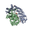
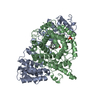
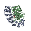
FPT is a dimer of the alpha and beta subunits. There is 1 alpha and 1 beta in the asymmetric unit and together these form the biological unit.
-
Components
-
Protein , 2 types, 2 molecules AB
#1: Protein
Proteinfarnesyltransferase/geranylgeranyltransferasetypeIalphasubunit / CAAX farnesyltransferase alpha subunit / Ras proteins prenyltransferase alpha / FTase-alpha / Type ...CAAX farnesyltransferase alpha subunit / Ras proteins prenyltransferase alpha / FTase-alpha / Type I protein geranyl-geranyltransferase alpha subunit / GGTase-I-alpha
Mass: 37691.344 Da / Num. of mol.: 1 Source method: isolated from a genetically manipulated source Source: (gene. exp.) Rattus norvegicus (Norway rat) / Gene: Fnta / Production host: Escherichia coli (E. coli) References: UniProt: Q04631, protein farnesyltransferase, protein geranylgeranyltransferase type I
In the structure databanks used in Yorodumi, some data are registered as the other names, "COVID-19 virus" and "2019-nCoV". Here are the details of the virus and the list of structure data.
Jan 31, 2019. EMDB accession codes are about to change! (news from PDBe EMDB page)
EMDB accession codes are about to change! (news from PDBe EMDB page)
The allocation of 4 digits for EMDB accession codes will soon come to an end. Whilst these codes will remain in use, new EMDB accession codes will include an additional digit and will expand incrementally as the available range of codes is exhausted. The current 4-digit format prefixed with “EMD-” (i.e. EMD-XXXX) will advance to a 5-digit format (i.e. EMD-XXXXX), and so on. It is currently estimated that the 4-digit codes will be depleted around Spring 2019, at which point the 5-digit format will come into force.
The EM Navigator/Yorodumi systems omit the EMD- prefix.
Related info.:Q: What is EMD? / ID/Accession-code notation in Yorodumi/EM Navigator
Yorodumi is a browser for structure data from EMDB, PDB, SASBDB, etc.
This page is also the successor to EM Navigator detail page, and also detail information page/front-end page for Omokage search.
The word "yorodu" (or yorozu) is an old Japanese word meaning "ten thousand". "mi" (miru) is to see.
Related info.:EMDB / PDB / SASBDB / Comparison of 3 databanks / Yorodumi Search / Aug 31, 2016. New EM Navigator & Yorodumi / Yorodumi Papers / Jmol/JSmol / Function and homology information / Changes in new EM Navigator and Yorodumi
 Movie
Movie Controller


 Open data
 Basic information
 Components
 Keywords
 Function and homology information
 X-RAY DIFFRACTION /
  Authors
 Citation
 Structure visualization
 Downloads & links
 Other downloads
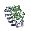


 PDBj
PDBj

 Assembly


 Mass: 65.409 Da / Num. of mol.: 1 / Source method: obtained synthetically / Formula: Zn
 Mass: 382.326 Da / Num. of mol.: 1 / Source method: obtained synthetically / Formula: C15H28O7P2
 Mass: 473.031 Da / Num. of mol.: 1 / Source method: obtained synthetically / Formula: C24H29ClN4O2S
 Sample preparation
 Processing