 Movie
Movie Controller

[English] 日本語
 Yorodumi
- PDB-3vgv: E134A mutant nucleoside diphosphate kinase derived from Halomonas... -
+ Open data

- Basic information
| Entry | Database: PDB / ID: 3vgv | ||||||
|---|---|---|---|---|---|---|---|
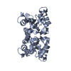
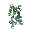
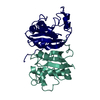
| Title | E134A mutant nucleoside diphosphate kinase derived from Halomonas sp. 593 | ||||||
 Components Components | Nucleoside diphosphate kinase | ||||||
 Keywords Keywords | TRANSFERASE / HALOPHILIC / KINASE / FERREDOXIN FOLD / ATP-BINDING / NUCLEOTIDE-BINDING | ||||||
| Function / homology |  Function and homology information Function and homology informationnucleoside-diphosphate kinase / UTP biosynthetic process / CTP biosynthetic process / nucleoside diphosphate kinase activity / GTP biosynthetic process / ATP binding / metal ion binding / cytoplasm Similarity search - Function | ||||||
| Biological species |  Halomonas (bacteria) Halomonas (bacteria) | ||||||
| Method |  X-RAY DIFFRACTION / SYNCHROTRON / MOLECULAR REPLACEMENT / Resolution: 2.5 Å X-RAY DIFFRACTION / SYNCHROTRON / MOLECULAR REPLACEMENT / Resolution: 2.5 Å | ||||||
 Authors Authors | Okazaki, N. / Yonezawa, Y. / Arai, S. / Matsumoto, F. / Tamada, T. / Tokunaga, H. / Ishibashi, M. / Tokunaga, M. / Kuroki, R. | ||||||
 Citation Citation | Journal: Protein Sci. / Year: 2012 Title: A structural mechanism for dimeric to tetrameric oligomer conversion in Halomonas sp. nucleoside diphosphate kinase Authors: Arai, S. / Yonezawa, Y. / Okazaki, N. / Matsumoto, F. / Tamada, T. / Tokunaga, H. / Ishibashi, M. / Blaber, M. / Tokunaga, M. / Kuroki, R. | ||||||
| History |
|
- Structure visualization
| Structure viewer | Molecule: MolmilJmol/JSmol |
|---|
- Downloads & links
-Download
| PDBx/mmCIF format | 3vgv.cif.gz | 405.1 KB | Display | PDBx/mmCIF format |
|---|---|---|---|---|
| PDB format | pdb3vgv.ent.gz | 336.2 KB | Display | PDB format |
| PDBx/mmJSON format | 3vgv.json.gz | Tree view | PDBx/mmJSON format | |
| Others |  Other downloads Other downloads |
-Validation report
| Arichive directory | https://data.pdbj.org/pub/pdb/validation_reports/vg/3vgvftp://data.pdbj.org/pub/pdb/validation_reports/vg/3vgv | HTTPS FTP |
|---|
-Related structure data
| Related structure data | 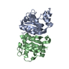 3vgsC  3vgtC  3vguC  1nhkS C: citing same article ( S: Starting model for refinement |
|---|---|
| Similar structure data |

-Links
 PDBj
PDBj- Assembly
| Deposited unit | 
| ||||||||
|---|---|---|---|---|---|---|---|---|---|
| 1 | 
| ||||||||
| 2 | 
| ||||||||
| 3 | 
| ||||||||
| 4 | 
| ||||||||
| 5 | 
| ||||||||
| 6 | 
| ||||||||
| 7 | 
| ||||||||
| 8 | 
| ||||||||
| Unit cell |
|
-Components
| #1: Protein | Mass: 15226.054 Da / Num. of mol.: 16 / Mutation: E134A Source method: isolated from a genetically manipulated source Source: (gene. exp.) Halomonas (bacteria) / Strain: 593 / Gene: NDK / Plasmid: PET / Production host: #2: Water | ChemComp-HOH / |  Mass: 18.015 Da / Num. of mol.: 208 / Source method: isolated from a natural source / Formula: H2O Mass: 18.015 Da / Num. of mol.: 208 / Source method: isolated from a natural source / Formula: H2O |
|---|
-Experimental details
-Experiment
| Experiment | Method: X-RAY DIFFRACTION / Number of used crystals: 1 |
|---|
- Sample preparation
| Crystal | Density Matthews: 2.4 Å3/Da / Density % sol: 48.72 % |
|---|---|
| Crystal grow | Temperature: 293 K / Method: vapor diffusion, sitting drop / pH: 5.6 Details: 0.2M CALCIUM ACETATE HYDRATE, 0.01M DITHIOTHREITOL (DTT), 0.1M SODIUM CACODYLATE TRIHYDRATE, 18% PEG 8000, pH 5.6, VAPOR DIFFUSION, SITTING DROP, temperature 293K |
-Data collection
| Diffraction | Mean temperature: 100 K |
|---|---|
| Diffraction source | Source: SYNCHROTRON / Site: Photon Factory  / Beamline: BL-6A / Wavelength: 0.978 / Beamline: BL-6A / Wavelength: 0.978 |
| Detector | Type: ADSC QUANTUM 4r / Detector: CCD / Date: Nov 12, 2008 |
| Radiation | Protocol: SINGLE WAVELENGTH / Monochromatic (M) / Laue (L): M / Scattering type: x-ray |
| Radiation wavelength | Wavelength: 0.978 Å / Relative weight: 1 |
| Reflection | Resolution: 2.5→42.95 Å / Num. obs: 73584 / % possible obs: 91.9 % / Redundancy: 2.5 % / Rmerge(I) obs: 0.1 / Net I/σ(I): 9.07 |
| Reflection shell | Resolution: 2.5→2.59 Å / Redundancy: 1.9 % / Rmerge(I) obs: 0.334 / Mean I/σ(I) obs: 1.9 / % possible all: 82.2 |
- Processing
| Software |
| ||||||||||||||||||||||||||||||||||||||||||||||||||||||||||||||||||||||||||||||||||||||||||||||||||||||||||||||||||||||||||||||||||||||||||||||||||||||||||||||||||||||||||
|---|---|---|---|---|---|---|---|---|---|---|---|---|---|---|---|---|---|---|---|---|---|---|---|---|---|---|---|---|---|---|---|---|---|---|---|---|---|---|---|---|---|---|---|---|---|---|---|---|---|---|---|---|---|---|---|---|---|---|---|---|---|---|---|---|---|---|---|---|---|---|---|---|---|---|---|---|---|---|---|---|---|---|---|---|---|---|---|---|---|---|---|---|---|---|---|---|---|---|---|---|---|---|---|---|---|---|---|---|---|---|---|---|---|---|---|---|---|---|---|---|---|---|---|---|---|---|---|---|---|---|---|---|---|---|---|---|---|---|---|---|---|---|---|---|---|---|---|---|---|---|---|---|---|---|---|---|---|---|---|---|---|---|---|---|---|---|---|---|---|---|---|
| Refinement | Method to determine structure: MOLECULAR REPLACEMENT Starting model: PDB ENTRY 1NHK Resolution: 2.5→42.95 Å / Cor.coef. Fo:Fc: 0.915 / Cor.coef. Fo:Fc free: 0.868 / SU B: 12.258 / SU ML: 0.263 / Cross valid method: THROUGHOUT / σ(F): 0 / ESU R Free: 0.345 / Stereochemistry target values: MAXIMUM LIKELIHOOD / Details: HYDROGENS HAVE BEEN ADDED IN THE RIDING
| ||||||||||||||||||||||||||||||||||||||||||||||||||||||||||||||||||||||||||||||||||||||||||||||||||||||||||||||||||||||||||||||||||||||||||||||||||||||||||||||||||||||||||
| Solvent computation | Ion probe radii: 0.8 Å / Shrinkage radii: 0.8 Å / VDW probe radii: 1.4 Å / Solvent model: MASK | ||||||||||||||||||||||||||||||||||||||||||||||||||||||||||||||||||||||||||||||||||||||||||||||||||||||||||||||||||||||||||||||||||||||||||||||||||||||||||||||||||||||||||
| Displacement parameters | Biso mean: 32.68 Å2
| ||||||||||||||||||||||||||||||||||||||||||||||||||||||||||||||||||||||||||||||||||||||||||||||||||||||||||||||||||||||||||||||||||||||||||||||||||||||||||||||||||||||||||
| Refinement step | Cycle: LAST / Resolution: 2.5→42.95 Å
| ||||||||||||||||||||||||||||||||||||||||||||||||||||||||||||||||||||||||||||||||||||||||||||||||||||||||||||||||||||||||||||||||||||||||||||||||||||||||||||||||||||||||||
| Refine LS restraints |
| ||||||||||||||||||||||||||||||||||||||||||||||||||||||||||||||||||||||||||||||||||||||||||||||||||||||||||||||||||||||||||||||||||||||||||||||||||||||||||||||||||||||||||
| LS refinement shell | Resolution: 2.5→2.56 Å / Total num. of bins used: 20
|
