 Movie
Movie Controller

[English] 日本語
 Yorodumi
- PDB-3vbi: Crystal Structure of AntD, an N-acyltransferase from Bacillus cer... -
+ Open data

- Basic information
| Entry | Database: PDB / ID: 3vbi | ||||||
|---|---|---|---|---|---|---|---|
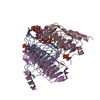
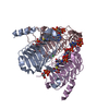
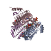
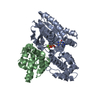
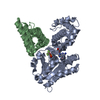
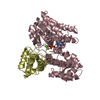
| Title | Crystal Structure of AntD, an N-acyltransferase from Bacillus cereus in complex with dTDP-4-amino-4,6-dideoxyglucose and Coenzyme A | ||||||
 Components Components | Galactoside O-acetyltransferase | ||||||
 Keywords Keywords | TRANSFERASE / anthrose / acylated sugar / left-handed beta helix / sugar N-acylation | ||||||
| Function / homology | Hexapeptide repeat proteins / UDP N-Acetylglucosamine Acyltransferase; domain 1 / 3 Solenoid / Mainly Beta / dTDP-4-amino-4,6-dideoxyglucose / BICARBONATE ION / COENZYME A / :  Function and homology information Function and homology information | ||||||
| Biological species |  | ||||||
| Method |  X-RAY DIFFRACTION / MOLECULAR REPLACEMENT / Resolution: 1.8 Å X-RAY DIFFRACTION / MOLECULAR REPLACEMENT / Resolution: 1.8 Å | ||||||
 Authors Authors | Kubiak, R.L. / Holden, H.M. | ||||||
 Citation Citation | Journal: Biochemistry / Year: 2012 Title: Structural Studies of AntD: An N-Acyltransferase Involved in the Biosynthesis of d-Anthrose. Authors: Kubiak, R.L. / Holden, H.M. | ||||||
| History |
|
- Structure visualization
| Structure viewer | Molecule: MolmilJmol/JSmol |
|---|
- Downloads & links
-Download
| PDBx/mmCIF format | 3vbi.cif.gz | 134.8 KB | Display | PDBx/mmCIF format |
|---|---|---|---|---|
| PDB format | pdb3vbi.ent.gz | 105.1 KB | Display | PDB format |
| PDBx/mmJSON format | 3vbi.json.gz | Tree view | PDBx/mmJSON format | |
| Others |  Other downloads Other downloads |
-Validation report
| Arichive directory | https://data.pdbj.org/pub/pdb/validation_reports/vb/3vbiftp://data.pdbj.org/pub/pdb/validation_reports/vb/3vbi | HTTPS FTP |
|---|
-Related structure data
| Related structure data |  3vbjC  3vbkC 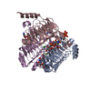 3vblC 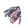 3vbmC 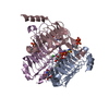 3vbnC  3vbpC C: citing same article ( |
|---|---|
| Similar structure data |


-Links
 PDBj
PDBj


- Assembly
| Deposited unit | 
| ||||||||
|---|---|---|---|---|---|---|---|---|---|
| 1 |
| ||||||||
| Unit cell |
|
-Components
-Protein , 1 types, 3 molecules ACE
| #1: Protein | Mass: 22752.293 Da / Num. of mol.: 3 Source method: isolated from a genetically manipulated source Source: (gene. exp.) References: UniProt: D7WGJ0, galactoside O-acetyltransferase |
|---|
-Non-polymers , 5 types, 308 molecules 


| #2: Chemical |  Mass: 767.534 Da / Num. of mol.: 3 / Source method: obtained synthetically / Formula: C21H36N7O16P3S Mass: 767.534 Da / Num. of mol.: 3 / Source method: obtained synthetically / Formula: C21H36N7O16P3S#3: Chemical |  Mass: 547.345 Da / Num. of mol.: 3 / Source method: obtained synthetically / Formula: C16H27N3O14P2 Mass: 547.345 Da / Num. of mol.: 3 / Source method: obtained synthetically / Formula: C16H27N3O14P2#4: Chemical |  Mass: 61.017 Da / Num. of mol.: 3 / Source method: obtained synthetically / Formula: CHO3 / Comment: pH buffer*YM Mass: 61.017 Da / Num. of mol.: 3 / Source method: obtained synthetically / Formula: CHO3 / Comment: pH buffer*YM#5: Chemical | ChemComp-CL / |  Mass: 35.453 Da / Num. of mol.: 1 / Source method: obtained synthetically / Formula: Cl Mass: 35.453 Da / Num. of mol.: 1 / Source method: obtained synthetically / Formula: Cl#6: Water | ChemComp-HOH / | Mass: 18.015 Da / Num. of mol.: 298 / Source method: isolated from a natural source / Formula: H2O |
|---|
-Experimental details
-Experiment
| Experiment | Method: X-RAY DIFFRACTION / Number of used crystals: 1 |
|---|
- Sample preparation
| Crystal | Density Matthews: 2.56 Å3/Da / Density % sol: 51.94 % |
|---|---|
| Crystal grow | Temperature: 298 K / Method: vapor diffusion, hanging drop / pH: 6 Details: 25% pentaerythritol ethoxylate (3/4 EO/OH), 2% isopropanol, pH 6.0, VAPOR DIFFUSION, HANGING DROP, temperature 298K |
-Data collection
| Diffraction | Mean temperature: 100 K |
|---|---|
| Diffraction source | Source: ROTATING ANODE / Type: RIGAKU RU200 / Wavelength: 1.54178 Å |
| Detector | Type: Bruker Platinum 135 / Detector: CCD / Date: May 9, 2011 / Details: Montel |
| Radiation | Monochromator: Nickel filter / Protocol: SINGLE WAVELENGTH / Monochromatic (M) / Laue (L): M / Scattering type: x-ray |
| Radiation wavelength | Wavelength: 1.54178 Å / Relative weight: 1 |
| Reflection | Resolution: 1.8→38.659 Å / Num. all: 63508 / Num. obs: 57751 / % possible obs: 90.9 % / Redundancy: 3.7 % / Rmerge(I) obs: 0.062 / Rsym value: 0.062 / Net I/σ(I): 11.8 |
| Reflection shell | Resolution: 1.8→1.9 Å / Redundancy: 1.6 % / Rmerge(I) obs: 0.249 / Mean I/σ(I) obs: 1.6 / Num. unique all: 7460 / Rsym value: 0.249 / % possible all: 78 |
- Processing
| Software |
| |||||||||||||||||||||||||||||||||||||||||||||||||||||||||||||||||
|---|---|---|---|---|---|---|---|---|---|---|---|---|---|---|---|---|---|---|---|---|---|---|---|---|---|---|---|---|---|---|---|---|---|---|---|---|---|---|---|---|---|---|---|---|---|---|---|---|---|---|---|---|---|---|---|---|---|---|---|---|---|---|---|---|---|---|
| Refinement | Method to determine structure: MOLECULAR REPLACEMENT Starting model: IN-HOUSE MIR MODEL Resolution: 1.8→38.659 Å / Cor.coef. Fo:Fc: 0.959 / Cor.coef. Fo:Fc free: 0.937 / SU B: 3.476 / SU ML: 0.102 / Cross valid method: THROUGHOUT / ESU R: 0.14 / ESU R Free: 0.138 / Stereochemistry target values: MAXIMUM LIKELIHOOD Details: HYDROGENS HAVE BEEN ADDED IN THE RIDING POSITIONS U VALUES : REFINED INDIVIDUALLY
| |||||||||||||||||||||||||||||||||||||||||||||||||||||||||||||||||
| Solvent computation | Ion probe radii: 0.8 Å / Shrinkage radii: 0.8 Å / VDW probe radii: 1.4 Å / Solvent model: MASK | |||||||||||||||||||||||||||||||||||||||||||||||||||||||||||||||||
| Displacement parameters | Biso mean: 27.478 Å2
| |||||||||||||||||||||||||||||||||||||||||||||||||||||||||||||||||
| Refinement step | Cycle: LAST / Resolution: 1.8→38.659 Å
| |||||||||||||||||||||||||||||||||||||||||||||||||||||||||||||||||
| Refine LS restraints |
| |||||||||||||||||||||||||||||||||||||||||||||||||||||||||||||||||
| LS refinement shell | Resolution: 1.8→1.846 Å / Total num. of bins used: 20
|
