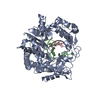
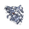
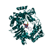
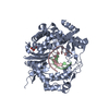
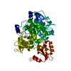
- PDB-3c7k: Molecular architecture of Galphao and the structural basis for RG... -
+
Open data
ID or keywords:
Loading...
-
Basic information
Entry
Database: PDB / ID: 3c7k
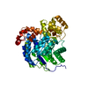
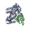
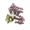
Title
Molecular architecture of Galphao and the structural basis for RGS16-mediated deactivation
Components
Guanine nucleotide-binding protein G(o) subunit alpha
Regulator of G-protein signaling 16
Keywords
SIGNALING PROTEIN / RGS / Galpha / AlF4 heterotrimeric G-protein GAP / GTP-binding / Lipoprotein / Myristate / Nucleotide-binding / Palmitate / Transducer / Phosphoprotein / Signal transduction inhibitor
Function / homology
Function and homology information
response to serotonin / G alpha (z) signalling events / potassium channel activator activity / Ca2+ pathway / G alpha (q) signalling events / G alpha (i) signalling events / mu-type opioid receptor binding / corticotropin-releasing hormone receptor 1 binding / GTPase activating protein binding / G protein-coupled dopamine receptor signaling pathway ...response to serotonin / G alpha (z) signalling events / potassium channel activator activity / Ca2+ pathway / G alpha (q) signalling events / G alpha (i) signalling events / mu-type opioid receptor binding / corticotropin-releasing hormone receptor 1 binding / GTPase activating protein binding / G protein-coupled dopamine receptor signaling pathway / negative regulation of G protein-coupled receptor signaling pathway / regulation of heart contraction / negative regulation of calcium ion transport / parallel fiber to Purkinje cell synapse / positive regulation of GTPase activity / negative regulation of insulin secretion / postsynaptic modulation of chemical synaptic transmission / adenylate cyclase-inhibiting serotonin receptor signaling pathway / G protein-coupled serotonin receptor binding / GTPase activator activity / locomotory behavior / GABA-ergic synapse / cytoplasmic side of plasma membrane / G-protein beta/gamma-subunit complex binding / adenylate cyclase-modulating G protein-coupled receptor signaling pathway / adenylate cyclase-inhibiting G protein-coupled receptor signaling pathway / myelin sheath / synaptic vesicle membrane / heterotrimeric G-protein complex / cell body / presynaptic membrane / G protein activity / Hydrolases; Acting on acid anhydrides; Acting on GTP to facilitate cellular and subcellular movement / postsynaptic membrane / G protein-coupled receptor signaling pathway / signaling receptor binding / GTPase activity / dendrite / GTP binding / glutamatergic synapse / membrane / metal ion binding / plasma membrane / cytoplasm Similarity search - Function
Regulator of G-protein Signalling 4; domain 1 - #10 / Regulator of G-protein Signalling 4; domain 1 / RGS, subdomain 1/3 / Regulator of G-protein Signalling 4, domain 2 / Regulator of G-protein Signalling 4; domain 2 / GI Alpha 1, domain 2-like / GI Alpha 1, domain 2-like / Regulator of G protein signaling domain / RGS domain / RGS domain profile. ...Regulator of G-protein Signalling 4; domain 1 - #10 / Regulator of G-protein Signalling 4; domain 1 / RGS, subdomain 1/3 / Regulator of G-protein Signalling 4, domain 2 / Regulator of G-protein Signalling 4; domain 2 / GI Alpha 1, domain 2-like / GI Alpha 1, domain 2-like / Regulator of G protein signaling domain / RGS domain / RGS domain profile. / Regulator of G protein signalling domain / RGS, subdomain 2 / RGS domain superfamily / G-protein alpha subunit, group I / G protein alpha subunit, helical insertion / G protein alpha subunit / Guanine nucleotide binding protein (G-protein), alpha subunit / G-protein alpha subunit / G-alpha domain profile. / P-loop containing nucleotide triphosphate hydrolases / Rossmann fold / P-loop containing nucleoside triphosphate hydrolase / Orthogonal Bundle / 3-Layer(aba) Sandwich / Mainly Alpha / Alpha Beta Similarity search - Domain/homology
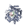
TETRAFLUOROALUMINATE ION / GUANOSINE-5'-DIPHOSPHATE / Guanine nucleotide-binding protein G(o) subunit alpha / Regulator of G protein signaling 16 Similarity search - Component
In the structure databanks used in Yorodumi, some data are registered as the other names, "COVID-19 virus" and "2019-nCoV". Here are the details of the virus and the list of structure data.
Jan 31, 2019. EMDB accession codes are about to change! (news from PDBe EMDB page)
EMDB accession codes are about to change! (news from PDBe EMDB page)
The allocation of 4 digits for EMDB accession codes will soon come to an end. Whilst these codes will remain in use, new EMDB accession codes will include an additional digit and will expand incrementally as the available range of codes is exhausted. The current 4-digit format prefixed with “EMD-” (i.e. EMD-XXXX) will advance to a 5-digit format (i.e. EMD-XXXXX), and so on. It is currently estimated that the 4-digit codes will be depleted around Spring 2019, at which point the 5-digit format will come into force.
The EM Navigator/Yorodumi systems omit the EMD- prefix.
Related info.:Q: What is EMD? / ID/Accession-code notation in Yorodumi/EM Navigator
Yorodumi is a browser for structure data from EMDB, PDB, SASBDB, etc.
This page is also the successor to EM Navigator detail page, and also detail information page/front-end page for Omokage search.
The word "yorodu" (or yorozu) is an old Japanese word meaning "ten thousand". "mi" (miru) is to see.
Related info.:EMDB / PDB / SASBDB / Comparison of 3 databanks / Yorodumi Search / Aug 31, 2016. New EM Navigator & Yorodumi / Yorodumi Papers / Jmol/JSmol / Function and homology information / Changes in new EM Navigator and Yorodumi
 Movie
Movie Controller

 Yorodumi
 Open data
 Basic information
 Components
 Keywords
 Function and homology information
 X-RAY DIFFRACTION /
  Authors
 Citation
 Structure visualization
 Downloads & links
 Other downloads
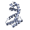
 PDBj
PDBj


 Assembly


 Mass: 24.305 Da / Num. of mol.: 2 / Source method: obtained synthetically / Formula: Mg
 Mass: 102.975 Da / Num. of mol.: 2 / Source method: obtained synthetically / Formula: AlF4
 Type: RNA linking / Mass: 443.201 Da / Num. of mol.: 2 / Source method: obtained synthetically / Formula: C10H15N5O11P2 / Comment: GDP, energy-carrying molecule*YM
 Sample preparation
 / Beamline: X25 / Wavelength: 1.15 Å
 Processing