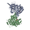
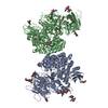
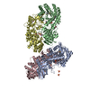
- PDB-3v8x: The crystal structure of transferrin binding protein A (TbpA) fro... -
+
Open data
ID or keywords:
Loading...
-
Basic information
Entry
Database: PDB / ID: 3v8x
Title
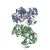
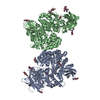
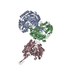
The crystal structure of transferrin binding protein A (TbpA) from Neisserial meningitidis serogroup B in complex with full length human transferrin
Components
Serotransferrin
Transferrin-binding protein 1
Keywords
MEMBRANE PROTEIN/METAL TRANSPORT / iron binding protein / transferrin binding protein A / iron binding/scavenging / MEMBRANE PROTEIN-METAL TRANSPORT complex
Function / homology
Function and homology information
ferric iron transmembrane transporter activity / siderophore transmembrane transport / iron chaperone activity / siderophore uptake transmembrane transporter activity / transferrin receptor binding / Transferrin endocytosis and recycling / basal part of cell / endocytic vesicle / clathrin-coated pit / ferric iron binding ...ferric iron transmembrane transporter activity / siderophore transmembrane transport / iron chaperone activity / siderophore uptake transmembrane transporter activity / transferrin receptor binding / Transferrin endocytosis and recycling / basal part of cell / endocytic vesicle / clathrin-coated pit / ferric iron binding / osteoclast differentiation / basal plasma membrane / Post-translational protein phosphorylation / cell outer membrane / iron ion transport / clathrin-coated endocytic vesicle membrane / HFE-transferrin receptor complex / regulation of protein stability / cellular response to iron ion / ferrous iron binding / Iron uptake and transport / positive regulation of receptor-mediated endocytosis / multicellular organismal-level iron ion homeostasis / recycling endosome / Regulation of Insulin-like Growth Factor (IGF) transport and uptake by Insulin-like Growth Factor Binding Proteins (IGFBPs) / late endosome / Platelet degranulation / Cargo recognition for clathrin-mediated endocytosis / positive regulation of proteasomal ubiquitin-dependent protein catabolic process / antibacterial humoral response / Clathrin-mediated endocytosis / cytoplasmic vesicle / secretory granule lumen / blood microparticle / vesicle / intracellular iron ion homeostasis / transmembrane transporter binding / early endosome / cell surface receptor signaling pathway / endosome membrane / apical plasma membrane / endoplasmic reticulum lumen / perinuclear region of cytoplasm / enzyme binding / cell surface / : / extracellular exosome / extracellular region / plasma membrane Similarity search - Function
In the structure databanks used in Yorodumi, some data are registered as the other names, "COVID-19 virus" and "2019-nCoV". Here are the details of the virus and the list of structure data.
Jan 31, 2019. EMDB accession codes are about to change! (news from PDBe EMDB page)
EMDB accession codes are about to change! (news from PDBe EMDB page)
The allocation of 4 digits for EMDB accession codes will soon come to an end. Whilst these codes will remain in use, new EMDB accession codes will include an additional digit and will expand incrementally as the available range of codes is exhausted. The current 4-digit format prefixed with “EMD-” (i.e. EMD-XXXX) will advance to a 5-digit format (i.e. EMD-XXXXX), and so on. It is currently estimated that the 4-digit codes will be depleted around Spring 2019, at which point the 5-digit format will come into force.
The EM Navigator/Yorodumi systems omit the EMD- prefix.
Related info.:Q: What is EMD? / ID/Accession-code notation in Yorodumi/EM Navigator
Yorodumi is a browser for structure data from EMDB, PDB, SASBDB, etc.
This page is also the successor to EM Navigator detail page, and also detail information page/front-end page for Omokage search.
The word "yorodu" (or yorozu) is an old Japanese word meaning "ten thousand". "mi" (miru) is to see.
Related info.:EMDB / PDB / SASBDB / Comparison of 3 databanks / Yorodumi Search / Aug 31, 2016. New EM Navigator & Yorodumi / Yorodumi Papers / Jmol/JSmol / Function and homology information / Changes in new EM Navigator and Yorodumi
 Movie
Movie Controller

 Yorodumi
 Open data
 Basic information
 Components
 Keywords
 Function and homology information
 Neisseria meningitidis serogroup B (bacteria)
 Homo sapiens (human)
 X-RAY DIFFRACTION /
  Authors
 Citation
 Structure visualization
 Downloads & links
 Other downloads
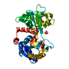


 PDBj
PDBj


 Assembly
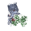


 Mass: 306.438 Da / Num. of mol.: 2 / Source method: obtained synthetically / Formula: C16H34O5 / Comment: C8E, detergent*YM
 Sample preparation
 / Beamline: 22-ID / Wavelength: 1 Å
 Processing