 Movie
Movie Controller

[English] 日本語
 Yorodumi
- PDB-3skp: The structure of apo-human transferrin C-lobe with bound sulfate ions -
+ Open data

- Basic information
| Entry | Database: PDB / ID: 3skp | ||||||
|---|---|---|---|---|---|---|---|
| Title | The structure of apo-human transferrin C-lobe with bound sulfate ions | ||||||
 Components Components | Serotransferrin | ||||||
 Keywords Keywords | METAL BINDING PROTEIN / transferrin / iron binding protein / transferrin receptor / TbpA / TbpB | ||||||
| Function / homology |  Function and homology information Function and homology informationiron chaperone activity / transferrin receptor binding / Transferrin endocytosis and recycling / basal part of cell / endocytic vesicle / clathrin-coated pit / osteoclast differentiation / ferric iron binding / basal plasma membrane / Post-translational protein phosphorylation ...iron chaperone activity / transferrin receptor binding / Transferrin endocytosis and recycling / basal part of cell / endocytic vesicle / clathrin-coated pit / osteoclast differentiation / ferric iron binding / basal plasma membrane / Post-translational protein phosphorylation / iron ion transport / clathrin-coated endocytic vesicle membrane / regulation of protein stability / HFE-transferrin receptor complex / cellular response to iron ion / ferrous iron binding / Iron uptake and transport / multicellular organismal-level iron ion homeostasis / positive regulation of receptor-mediated endocytosis / recycling endosome / Regulation of Insulin-like Growth Factor (IGF) transport and uptake by Insulin-like Growth Factor Binding Proteins (IGFBPs) / Platelet degranulation / late endosome / Cargo recognition for clathrin-mediated endocytosis / positive regulation of proteasomal ubiquitin-dependent protein catabolic process / antibacterial humoral response / Clathrin-mediated endocytosis / cytoplasmic vesicle / secretory granule lumen / blood microparticle / vesicle / intracellular iron ion homeostasis / transmembrane transporter binding / early endosome / cell surface receptor signaling pathway / apical plasma membrane / endosome membrane / endoplasmic reticulum lumen / perinuclear region of cytoplasm / enzyme binding / cell surface / : / extracellular exosome / extracellular region / plasma membrane Similarity search - Function | ||||||
| Biological species |  Homo sapiens (human) Homo sapiens (human) | ||||||
| Method |  X-RAY DIFFRACTION / SYNCHROTRON / MOLECULAR REPLACEMENT / Resolution: 1.7 Å X-RAY DIFFRACTION / SYNCHROTRON / MOLECULAR REPLACEMENT / Resolution: 1.7 Å | ||||||
 Authors Authors | Noinaj, N. / Steere, A.N. / Mason, A.B. / Buchanan, S.K. | ||||||
 Citation Citation | Journal: Nature / Year: 2012 Title: Structural basis for iron piracy by pathogenic Neisseria. Authors: Noinaj, N. / Easley, N.C. / Oke, M. / Mizuno, N. / Gumbart, J. / Boura, E. / Steere, A.N. / Zak, O. / Aisen, P. / Tajkhorshid, E. / Evans, R.W. / Gorringe, A.R. / Mason, A.B. / Steven, A.C. / Buchanan, S.K. | ||||||
| History |
|
- Structure visualization
| Structure viewer | Molecule: MolmilJmol/JSmol |
|---|
- Downloads & links
-Download
| PDBx/mmCIF format | 3skp.cif.gz | 152 KB | Display | PDBx/mmCIF format |
|---|---|---|---|---|
| PDB format | pdb3skp.ent.gz | 119.1 KB | Display | PDB format |
| PDBx/mmJSON format | 3skp.json.gz | Tree view | PDBx/mmJSON format | |
| Others |  Other downloads Other downloads |
-Validation report
| Arichive directory | https://data.pdbj.org/pub/pdb/validation_reports/sk/3skpftp://data.pdbj.org/pub/pdb/validation_reports/sk/3skp | HTTPS FTP |
|---|
-Related structure data
| Related structure data |  3v83C  3v89C  3v8uC  3v8xC  2havS S: Starting model for refinement C: citing same article ( |
|---|---|
| Similar structure data |


-Links
 PDBj
PDBj


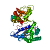
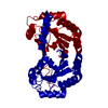
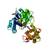
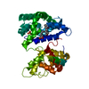
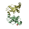
- Assembly
| Deposited unit | 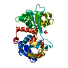
| ||||||||
|---|---|---|---|---|---|---|---|---|---|
| 1 |
| ||||||||
| 2 | 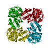
| ||||||||
| 3 | 
| ||||||||
| Unit cell |
| ||||||||
| Components on special symmetry positions |
|
-Components
| #1: Protein | Mass: 38015.176 Da / Num. of mol.: 1 / Fragment: iron binding protein Source method: isolated from a genetically manipulated source Source: (gene. exp.) Homo sapiens (human) / Gene: TF, PRO1400 / Cell (production host): baby hamster kidney (BHK) cells / Production host:  Cricetinae (hamsters) / References: UniProt: P02787 Cricetinae (hamsters) / References: UniProt: P02787 | ||||||
|---|---|---|---|---|---|---|---|
| #2: Chemical | ChemComp-SO4 /   Mass: 96.063 Da / Num. of mol.: 8 / Source method: obtained synthetically / Formula: SO4 Mass: 96.063 Da / Num. of mol.: 8 / Source method: obtained synthetically / Formula: SO4#3: Water | ChemComp-HOH / |  Mass: 18.015 Da / Num. of mol.: 370 / Source method: isolated from a natural source / Formula: H2O Mass: 18.015 Da / Num. of mol.: 370 / Source method: isolated from a natural source / Formula: H2OHas protein modification | Y | Sequence details | THIS IS A NATURAL VARIANT ACCORDING TO UNIPROT DATABASE. | |
-Experimental details
-Experiment
| Experiment | Method: X-RAY DIFFRACTION / Number of used crystals: 1 |
|---|
- Sample preparation
| Crystal | Density Matthews: 3.08 Å3/Da / Density % sol: 60.11 % |
|---|---|
| Crystal grow | Temperature: 294 K / Method: vapor diffusion, hanging drop / pH: 7.5 Details: 0.2 M potassium sulfate, 2.2 M ammonium sulfate, pH 7.5, VAPOR DIFFUSION, HANGING DROP, temperature 294K |
-Data collection
| Diffraction | Mean temperature: 100 K |
|---|---|
| Diffraction source | Source: SYNCHROTRON / Site: APS  / Beamline: 22-ID / Wavelength: 1 Å / Beamline: 22-ID / Wavelength: 1 Å |
| Detector | Type: MARMOSAIC 300 mm CCD / Detector: CCD / Date: Oct 15, 2010 |
| Radiation | Monochromator: double crystal / Protocol: SINGLE WAVELENGTH / Monochromatic (M) / Laue (L): M / Scattering type: x-ray |
| Radiation wavelength | Wavelength: 1 Å / Relative weight: 1 |
| Reflection | Resolution: 1.7→36.27 Å / Num. all: 52589 / Num. obs: 52589 / % possible obs: 99.9 % / Observed criterion σ(F): 1 / Observed criterion σ(I): 1 / Redundancy: 7 % / Biso Wilson estimate: 20.76 Å2 / Rsym value: 0.084 / Net I/σ(I): 31.2 |
| Reflection shell | Resolution: 1.7→1.76 Å / Redundancy: 6.3 % / Mean I/σ(I) obs: 2.2 / Rsym value: 0.832 / % possible all: 99.9 |
- Processing
| Software |
| ||||||||||||||||||||||||||||||||||||||||||||||||||||||||||||||||||||||||||||||||||||||||||||||||||||||||||||||||||||||||||||||||||||||||||||
|---|---|---|---|---|---|---|---|---|---|---|---|---|---|---|---|---|---|---|---|---|---|---|---|---|---|---|---|---|---|---|---|---|---|---|---|---|---|---|---|---|---|---|---|---|---|---|---|---|---|---|---|---|---|---|---|---|---|---|---|---|---|---|---|---|---|---|---|---|---|---|---|---|---|---|---|---|---|---|---|---|---|---|---|---|---|---|---|---|---|---|---|---|---|---|---|---|---|---|---|---|---|---|---|---|---|---|---|---|---|---|---|---|---|---|---|---|---|---|---|---|---|---|---|---|---|---|---|---|---|---|---|---|---|---|---|---|---|---|---|---|---|
| Refinement | Method to determine structure: MOLECULAR REPLACEMENT Starting model: PDB entry 2HAV Resolution: 1.7→29.563 Å / SU ML: 0.2 / Cross valid method: THROUGHOUT / σ(F): 0 / Phase error: 18.25 / Stereochemistry target values: ML
| ||||||||||||||||||||||||||||||||||||||||||||||||||||||||||||||||||||||||||||||||||||||||||||||||||||||||||||||||||||||||||||||||||||||||||||
| Solvent computation | Shrinkage radii: 0.95 Å / VDW probe radii: 1.2 Å / Solvent model: FLAT BULK SOLVENT MODEL / Bsol: 37.385 Å2 / ksol: 0.357 e/Å3 | ||||||||||||||||||||||||||||||||||||||||||||||||||||||||||||||||||||||||||||||||||||||||||||||||||||||||||||||||||||||||||||||||||||||||||||
| Displacement parameters |
| ||||||||||||||||||||||||||||||||||||||||||||||||||||||||||||||||||||||||||||||||||||||||||||||||||||||||||||||||||||||||||||||||||||||||||||
| Refinement step | Cycle: LAST / Resolution: 1.7→29.563 Å
| ||||||||||||||||||||||||||||||||||||||||||||||||||||||||||||||||||||||||||||||||||||||||||||||||||||||||||||||||||||||||||||||||||||||||||||
| Refine LS restraints |
| ||||||||||||||||||||||||||||||||||||||||||||||||||||||||||||||||||||||||||||||||||||||||||||||||||||||||||||||||||||||||||||||||||||||||||||
| LS refinement shell |
| ||||||||||||||||||||||||||||||||||||||||||||||||||||||||||||||||||||||||||||||||||||||||||||||||||||||||||||||||||||||||||||||||||||||||||||
| Refinement TLS params. | Method: refined / Refine-ID: X-RAY DIFFRACTION
| ||||||||||||||||||||||||||||||||||||||||||||||||||||||||||||||||||||||||||||||||||||||||||||||||||||||||||||||||||||||||||||||||||||||||||||
| Refinement TLS group |
|
