 Movie
Movie Controller


+ Open data

- Basic information
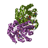
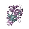
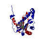
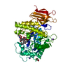
| Entry | Database: PDB / ID: 3v6e | ||||||
|---|---|---|---|---|---|---|---|
| Title | Crystal Structure of USP2 and a mutant form of Ubiquitin | ||||||
 Components Components |
| ||||||
 Keywords Keywords | hydrolase/signaling protein / Structural Genomics / Structural Genomics Consortium / SGC / ubiquitin / protease / hydrolase-signaling protein complex | ||||||
| Function / homology |  Function and homology information Function and homology informationcircadian behavior / TNFR1-induced proapoptotic signaling / muscle organ development / entrainment of circadian clock by photoperiod / locomotor rhythm / protein deubiquitination / Maturation of protein E / Maturation of protein E / ER Quality Control Compartment (ERQC) / Myoclonic epilepsy of Lafora ...circadian behavior / TNFR1-induced proapoptotic signaling / muscle organ development / entrainment of circadian clock by photoperiod / locomotor rhythm / protein deubiquitination / Maturation of protein E / Maturation of protein E / ER Quality Control Compartment (ERQC) / Myoclonic epilepsy of Lafora / positive regulation of mitotic cell cycle / FLT3 signaling by CBL mutants / IRAK2 mediated activation of TAK1 complex / Prevention of phagosomal-lysosomal fusion / Alpha-protein kinase 1 signaling pathway / Glycogen synthesis / IRAK1 recruits IKK complex / IRAK1 recruits IKK complex upon TLR7/8 or 9 stimulation / Endosomal Sorting Complex Required For Transport (ESCRT) / Membrane binding and targetting of GAG proteins / Negative regulation of FLT3 / Regulation of TBK1, IKKε (IKBKE)-mediated activation of IRF3, IRF7 / PTK6 Regulates RTKs and Their Effectors AKT1 and DOK1 / Regulation of TBK1, IKKε-mediated activation of IRF3, IRF7 upon TLR3 ligation / IRAK2 mediated activation of TAK1 complex upon TLR7/8 or 9 stimulation / Constitutive Signaling by NOTCH1 HD Domain Mutants / NOTCH2 Activation and Transmission of Signal to the Nucleus / TICAM1,TRAF6-dependent induction of TAK1 complex / TICAM1-dependent activation of IRF3/IRF7 / APC/C:Cdc20 mediated degradation of Cyclin B / Regulation of FZD by ubiquitination / Downregulation of ERBB4 signaling / APC-Cdc20 mediated degradation of Nek2A / p75NTR recruits signalling complexes / InlA-mediated entry of Listeria monocytogenes into host cells / TRAF6 mediated IRF7 activation in TLR7/8 or 9 signaling / NF-kB is activated and signals survival / TRAF6-mediated induction of TAK1 complex within TLR4 complex / Regulation of pyruvate metabolism / cyclin binding / Pexophagy / Regulation of innate immune responses to cytosolic DNA / NRIF signals cell death from the nucleus / Downregulation of ERBB2:ERBB3 signaling / Regulation of PTEN localization / VLDLR internalisation and degradation / regulation of signal transduction by p53 class mediator / Activated NOTCH1 Transmits Signal to the Nucleus / Synthesis of active ubiquitin: roles of E1 and E2 enzymes / Translesion synthesis by REV1 / TICAM1, RIP1-mediated IKK complex recruitment / Regulation of BACH1 activity / Translesion synthesis by POLK / InlB-mediated entry of Listeria monocytogenes into host cell / JNK (c-Jun kinases) phosphorylation and activation mediated by activated human TAK1 / MAP3K8 (TPL2)-dependent MAPK1/3 activation / Activation of IRF3, IRF7 mediated by TBK1, IKKε (IKBKE) / Downregulation of TGF-beta receptor signaling / Translesion synthesis by POLI / Josephin domain DUBs / IKK complex recruitment mediated by RIP1 / Gap-filling DNA repair synthesis and ligation in GG-NER / PINK1-PRKN Mediated Mitophagy / TGF-beta receptor signaling in EMT (epithelial to mesenchymal transition) / Regulation of activated PAK-2p34 by proteasome mediated degradation / TNFR1-induced NF-kappa-B signaling pathway / TCF dependent signaling in response to WNT / Regulation of NF-kappa B signaling / activated TAK1 mediates p38 MAPK activation / Autodegradation of Cdh1 by Cdh1:APC/C / APC/C:Cdc20 mediated degradation of Securin / circadian regulation of gene expression / Asymmetric localization of PCP proteins / Regulation of signaling by CBL / N-glycan trimming in the ER and Calnexin/Calreticulin cycle / Ubiquitin-dependent degradation of Cyclin D / NOTCH3 Activation and Transmission of Signal to the Nucleus / Negative regulators of DDX58/IFIH1 signaling / Fanconi Anemia Pathway / Peroxisomal protein import / SCF-beta-TrCP mediated degradation of Emi1 / NIK-->noncanonical NF-kB signaling / Negative regulation of FGFR3 signaling / AUF1 (hnRNP D0) binds and destabilizes mRNA / TNFR2 non-canonical NF-kB pathway / Deactivation of the beta-catenin transactivating complex / Stabilization of p53 / Assembly of the pre-replicative complex / Negative regulation of FGFR2 signaling / Vpu mediated degradation of CD4 / Negative regulation of FGFR4 signaling / Downregulation of SMAD2/3:SMAD4 transcriptional activity / Negative regulation of FGFR1 signaling / Cdc20:Phospho-APC/C mediated degradation of Cyclin A / Termination of translesion DNA synthesis / EGFR downregulation / Regulation of TNFR1 signaling / Dectin-1 mediated noncanonical NF-kB signaling / Degradation of DVL / Degradation of CRY and PER proteins Similarity search - Function | ||||||
| Biological species |  Homo sapiens (human) Homo sapiens (human) | ||||||
| Method |  X-RAY DIFFRACTION / MOLECULAR REPLACEMENT / molecular replacement / Resolution: 2.1 Å X-RAY DIFFRACTION / MOLECULAR REPLACEMENT / molecular replacement / Resolution: 2.1 Å | ||||||
 Authors Authors | Neculai, M. / Ernst, A. / Sidhu, S. / Arrowsmith, C.H. / Edwards, A.M. / Bountra, C. / Weigelt, J. / Dhe-Paganon, S. / Structural Genomics Consortium (SGC) | ||||||
 Citation Citation | Journal: Science / Year: 2013 Title: A strategy for modulation of enzymes in the ubiquitin system. Authors: Ernst, A. / Avvakumov, G. / Tong, J. / Fan, Y. / Zhao, Y. / Alberts, P. / Persaud, A. / Walker, J.R. / Neculai, A.M. / Neculai, D. / Vorobyov, A. / Garg, P. / Beatty, L. / Chan, P.K. / ...Authors: Ernst, A. / Avvakumov, G. / Tong, J. / Fan, Y. / Zhao, Y. / Alberts, P. / Persaud, A. / Walker, J.R. / Neculai, A.M. / Neculai, D. / Vorobyov, A. / Garg, P. / Beatty, L. / Chan, P.K. / Juang, Y.C. / Landry, M.C. / Yeh, C. / Zeqiraj, E. / Karamboulas, K. / Allali-Hassani, A. / Vedadi, M. / Tyers, M. / Moffat, J. / Sicheri, F. / Pelletier, L. / Durocher, D. / Raught, B. / Rotin, D. / Yang, J. / Moran, M.F. / Dhe-Paganon, S. / Sidhu, S.S. | ||||||
| History |
|
- Structure visualization
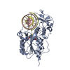
| Structure viewer | Molecule: MolmilJmol/JSmol |
|---|
- Downloads & links
-Download
| PDBx/mmCIF format | 3v6e.cif.gz | 105.3 KB | Display | PDBx/mmCIF format |
|---|---|---|---|---|
| PDB format | pdb3v6e.ent.gz | 78.9 KB | Display | PDB format |
| PDBx/mmJSON format | 3v6e.json.gz | Tree view | PDBx/mmJSON format | |
| Others |  Other downloads Other downloads |
-Validation report
| Arichive directory | https://data.pdbj.org/pub/pdb/validation_reports/v6/3v6eftp://data.pdbj.org/pub/pdb/validation_reports/v6/3v6e | HTTPS FTP |
|---|
-Related structure data
| Related structure data | 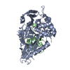 3i3tC 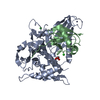 3mtnC  3n3kC  4i6lC  2hd5S S: Starting model for refinement C: citing same article ( |
|---|---|
| Similar structure data |
-Links
 PDBj
PDBj


- Assembly
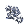
| Deposited unit | 
| ||||||||
|---|---|---|---|---|---|---|---|---|---|
| 1 |
| ||||||||
| Unit cell |
|
-Components
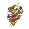
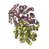
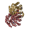
-Protein , 2 types, 2 molecules AB
| #1: Protein | Mass: 42171.719 Da / Num. of mol.: 1 Source method: isolated from a genetically manipulated source Source: (gene. exp.) Homo sapiens (human) / Gene: UBP41, usp02.258.605, USP2 / Plasmid: PET28LIC / Production host:  |
|---|---|
| #2: Protein | Mass: 10343.754 Da / Num. of mol.: 1 / Mutation: G76S, K82N, T85S, T88H Source method: isolated from a genetically manipulated source Source: (gene. exp.) Homo sapiens (human) / Gene: UBC / Plasmid: PET53 / Production host: |
-Non-polymers , 4 types, 201 molecules 


| #3: Chemical | ChemComp-ZN /  Mass: 65.409 Da / Num. of mol.: 1 / Source method: obtained synthetically / Formula: Zn Mass: 65.409 Da / Num. of mol.: 1 / Source method: obtained synthetically / Formula: Zn | ||||
|---|---|---|---|---|---|
| #4: Chemical |  Mass: 35.453 Da / Num. of mol.: 2 / Source method: obtained synthetically / Formula: Cl Mass: 35.453 Da / Num. of mol.: 2 / Source method: obtained synthetically / Formula: Cl#5: Chemical | ChemComp-GOL / |  Mass: 92.094 Da / Num. of mol.: 1 / Source method: obtained synthetically / Formula: C3H8O3 Mass: 92.094 Da / Num. of mol.: 1 / Source method: obtained synthetically / Formula: C3H8O3#6: Water | ChemComp-HOH / | Mass: 18.015 Da / Num. of mol.: 197 / Source method: isolated from a natural source / Formula: H2O |
-Experimental details
-Experiment
| Experiment | Method: X-RAY DIFFRACTION / Number of used crystals: 1 |
|---|
- Sample preparation
| Crystal | Density Matthews: 2.03 Å3/Da / Density % sol: 39.49 % |
|---|---|
| Crystal grow | Temperature: 293 K / Method: vapor diffusion, hanging drop / pH: 7 Details: 21% PEG3350, 0.2 M Calcium Chloride,0.1 Bis-Tris pH 7.0, glycerol, VAPOR DIFFUSION, HANGING DROP, temperature 293K |
-Data collection
| Diffraction | Mean temperature: 100 K |
|---|---|
| Diffraction source | Source: ROTATING ANODE / Type: RIGAKU FR-E+ SUPERBRIGHT / Wavelength: 1.54178 Å |
| Detector | Type: RIGAKU RAXIS IV++ / Detector: IMAGE PLATE / Date: Nov 29, 2011 |
| Radiation | Protocol: SINGLE WAVELENGTH / Monochromatic (M) / Laue (L): M / Scattering type: x-ray |
| Radiation wavelength | Wavelength: 1.54178 Å / Relative weight: 1 |
| Reflection | Resolution: 2→85 Å / Num. all: 29672 / Num. obs: 29660 / % possible obs: 100 % / Observed criterion σ(F): 0 / Observed criterion σ(I): 0 / Redundancy: 7.04 % / Rsym value: 0.1589 / Net I/σ(I): 11.5 |
| Reflection shell | Resolution: 2→2.04 Å / Redundancy: 6.98 % / Mean I/σ(I) obs: 3.01 / Num. unique all: 1252 / Rsym value: 0.5588 / % possible all: 99.9 |
-Phasing
| Phasing | Method: molecular replacement |
|---|
- Processing
| Software |
| |||||||||||||||||||||||||||||||||||||||||||||||||||||||||||||||||
|---|---|---|---|---|---|---|---|---|---|---|---|---|---|---|---|---|---|---|---|---|---|---|---|---|---|---|---|---|---|---|---|---|---|---|---|---|---|---|---|---|---|---|---|---|---|---|---|---|---|---|---|---|---|---|---|---|---|---|---|---|---|---|---|---|---|---|
| Refinement | Method to determine structure: MOLECULAR REPLACEMENT Starting model: pdb entry 2HD5 Resolution: 2.1→42.06 Å / Cor.coef. Fo:Fc: 0.941 / Cor.coef. Fo:Fc free: 0.9 / WRfactor Rfree: 0.2018 / WRfactor Rwork: 0.15 / Occupancy max: 1 / Occupancy min: 0.2 / FOM work R set: 0.8767 / SU B: 4.375 / SU ML: 0.119 / SU R Cruickshank DPI: 0.2261 / SU Rfree: 0.1937 / Cross valid method: THROUGHOUT / σ(F): 0 / ESU R: 0.226 / ESU R Free: 0.194 / Stereochemistry target values: MAXIMUM LIKELIHOOD Details: HYDROGENS HAVE BEEN ADDED IN THE RIDING POSITIONS U VALUES : REFINED INDIVIDUALLY
| |||||||||||||||||||||||||||||||||||||||||||||||||||||||||||||||||
| Solvent computation | Ion probe radii: 0.8 Å / Shrinkage radii: 0.8 Å / VDW probe radii: 1.4 Å / Solvent model: MASK | |||||||||||||||||||||||||||||||||||||||||||||||||||||||||||||||||
| Displacement parameters | Biso max: 45.81 Å2 / Biso mean: 15.1054 Å2 / Biso min: 2 Å2
| |||||||||||||||||||||||||||||||||||||||||||||||||||||||||||||||||
| Refinement step | Cycle: LAST / Resolution: 2.1→42.06 Å
| |||||||||||||||||||||||||||||||||||||||||||||||||||||||||||||||||
| Refine LS restraints |
| |||||||||||||||||||||||||||||||||||||||||||||||||||||||||||||||||
| LS refinement shell | Resolution: 2.1→2.154 Å / Total num. of bins used: 20
|
