 Movie
Movie Controller

[English] 日本語
 Yorodumi
- PDB-1jjk: Selenomethionine Substitution of Orotidine-5'-monophosphate Decar... -
+ Open data

- Basic information
| Entry | Database: PDB / ID: 1jjk | ||||||
|---|---|---|---|---|---|---|---|
| Title | Selenomethionine Substitution of Orotidine-5'-monophosphate Decarboxylase from E. coli Causes a Change in Crystal Contacts and Space Group | ||||||
 Components Components | OROTIDINE 5'-PHOSPHATE DECARBOXYLASE | ||||||
 Keywords Keywords | LYASE / alpha-beta-barrel / protein-inhibitor complex / homodimer | ||||||
| Function / homology |  Function and homology information Function and homology informationnucleobase-containing small molecule interconversion / carboxy-lyase activity / orotidine-5'-phosphate decarboxylase / orotidine-5'-phosphate decarboxylase activity / 'de novo' UMP biosynthetic process / 'de novo' pyrimidine nucleobase biosynthetic process / cytosol / cytoplasm Similarity search - Function | ||||||
| Biological species |  | ||||||
| Method |  X-RAY DIFFRACTION / SYNCHROTRON / MAD / Resolution: 3 Å X-RAY DIFFRACTION / SYNCHROTRON / MAD / Resolution: 3 Å | ||||||
 Authors Authors | Poulsen, J.-C.N. / Harris, P. / Jensen, K.F. / Larsen, S. | ||||||
 Citation Citation | Journal: Acta Crystallogr.,Sect.D / Year: 2001 Title: Selenomethionine substitution of orotidine-5'-monophosphate decarboxylase causes a change in crystal contacts and space group. Authors: Poulsen, J.C. / Harris, P. / Jensen, K.F. / Larsen, S. #1: Journal: Biochemistry / Year: 2000Title: Structural Basis for the Catalytic Mechanism of a Proficient Enzyme: Orotidine 5'-monophosphate Decarboxylase Authors: Harris, P. / Poulsen, J.-C.N. / Jensen, K.F. / Larsen, S. | ||||||
| History |
|
- Structure visualization
| Structure viewer | Molecule: MolmilJmol/JSmol |
|---|
- Downloads & links
-Download
| PDBx/mmCIF format | 1jjk.cif.gz | 688.7 KB | Display | PDBx/mmCIF format |
|---|---|---|---|---|
| PDB format | pdb1jjk.ent.gz | 575.3 KB | Display | PDB format |
| PDBx/mmJSON format | 1jjk.json.gz | Tree view | PDBx/mmJSON format | |
| Others |  Other downloads Other downloads |
-Validation report
| Arichive directory | https://data.pdbj.org/pub/pdb/validation_reports/jj/1jjkftp://data.pdbj.org/pub/pdb/validation_reports/jj/1jjk | HTTPS FTP |
|---|
-Related structure data
| Related structure data | |
|---|---|
| Similar structure data |


-Links
 PDBj
PDBj
- Assembly
| Deposited unit | 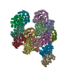
| ||||||||
|---|---|---|---|---|---|---|---|---|---|
| 1 | 
| ||||||||
| 2 | 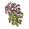
| ||||||||
| 3 | 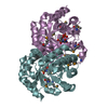
| ||||||||
| 4 | 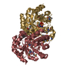
| ||||||||
| 5 | 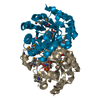
| ||||||||
| 6 | 
| ||||||||
| 7 | 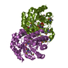
| ||||||||
| 8 | 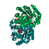
| ||||||||
| Unit cell |
| ||||||||
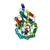
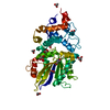
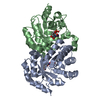
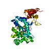
| Details | The biological assembly is a dimer constructed like AB, CD, EF, GH, IJ, KL, MN or OP |
-Components
| #1: Protein | Mass: 26800.279 Da / Num. of mol.: 16 Source method: isolated from a genetically manipulated source Source: (gene. exp.) References: UniProt: P08244, orotidine-5'-phosphate decarboxylase #2: Chemical | ChemComp-BMP /   Type: RNA linking / Mass: 340.181 Da / Num. of mol.: 16 / Source method: obtained synthetically / Formula: C9H13N2O10P Type: RNA linking / Mass: 340.181 Da / Num. of mol.: 16 / Source method: obtained synthetically / Formula: C9H13N2O10PHas protein modification | Y | |
|---|
-Experimental details
-Experiment
| Experiment | Method: X-RAY DIFFRACTION / Number of used crystals: 1 |
|---|
- Sample preparation
| Crystal | Density Matthews: 2.1 Å3/Da / Density % sol: 41.32 % | ||||||||||||||||||||||||||||||
|---|---|---|---|---|---|---|---|---|---|---|---|---|---|---|---|---|---|---|---|---|---|---|---|---|---|---|---|---|---|---|---|
| Crystal grow | Temperature: 290 K / Method: vapor diffusion, hanging drop / pH: 7 Details: PEG 8000, magnesium chloride, pH 7.0, VAPOR DIFFUSION, HANGING DROP, temperature 290K | ||||||||||||||||||||||||||||||
| Crystal grow | *PLUS Temperature: 291 K | ||||||||||||||||||||||||||||||
| Components of the solutions | *PLUS
|
-Data collection
| Diffraction | Mean temperature: 100 K | ||||||||||||
|---|---|---|---|---|---|---|---|---|---|---|---|---|---|
| Diffraction source | Source: SYNCHROTRON / Site: ESRF  / Beamline: BM14 / Wavelength: 0.9787, 0.9789, 0.8856 / Beamline: BM14 / Wavelength: 0.9787, 0.9789, 0.8856 | ||||||||||||
| Detector | Type: MARRESEARCH / Detector: IMAGE PLATE / Date: Apr 9, 1999 | ||||||||||||
| Radiation | Monochromator: Si(111) monochromator / Protocol: MAD / Monochromatic (M) / Laue (L): M / Scattering type: x-ray | ||||||||||||
| Radiation wavelength |
| ||||||||||||
| Reflection | Resolution: 3→30 Å / Num. all: 64921 / Num. obs: 64921 / % possible obs: 92.3 % / Observed criterion σ(F): 0 / Observed criterion σ(I): 0 / Redundancy: 5.1 % / Biso Wilson estimate: 47 Å2 / Rmerge(I) obs: 0.14 / Net I/σ(I): 3.1 | ||||||||||||
| Reflection shell | Resolution: 3→3.16 Å / Redundancy: 4.8 % / Rmerge(I) obs: 0.315 / Mean I/σ(I) obs: 1.3 / Num. unique all: 9615 / % possible all: 93.7 | ||||||||||||
| Reflection | *PLUS Lowest resolution: 30 Å / Num. measured all: 333946 |
- Processing
| Software |
| |||||||||||||||||||||||||
|---|---|---|---|---|---|---|---|---|---|---|---|---|---|---|---|---|---|---|---|---|---|---|---|---|---|---|
| Refinement | Method to determine structure: MAD / Resolution: 3→30 Å / σ(F): 0 / σ(I): 0 / Stereochemistry target values: Engh & huber
| |||||||||||||||||||||||||
| Refinement step | Cycle: LAST / Resolution: 3→30 Å
| |||||||||||||||||||||||||
| Refine LS restraints |
| |||||||||||||||||||||||||
| LS refinement shell | Resolution: 3→3.02 Å
| |||||||||||||||||||||||||
| Software | *PLUS Name: CNS / Version: 0.9 / Classification: refinement | |||||||||||||||||||||||||
| Refinement | *PLUS Highest resolution: 3 Å / Lowest resolution: 30 Å / σ(F): 0 | |||||||||||||||||||||||||
| Solvent computation | *PLUS | |||||||||||||||||||||||||
| Displacement parameters | *PLUS | |||||||||||||||||||||||||
| Refine LS restraints | *PLUS Type: c_angle_deg / Dev ideal: 1.3 | |||||||||||||||||||||||||
| LS refinement shell | *PLUS Highest resolution: 3 Å / Rfactor Rfree: 0.387 / Rfactor Rwork: 0.423 |
