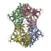
[English] 日本語
Yorodumi
- PDB-3ute: Crystal structure of Aspergillus fumigatus UDP galactopyranose mu... -

+
Open data


ID or keywords:

Loading...

-
Basic information

Entry
Database: PDB / ID: 3ute
TitleCrystal structure of Aspergillus fumigatus UDP galactopyranose mutase sulfate complex
ComponentsUDP-galactopyranose mutase
KeywordsISOMERASE / Nucleotide binding / Mutase / Flavin adenine dinucleotide binding
Function / homology
Function and homology information


UDP-galactopyranose mutase / UDP-galactopyranose mutase activity / nucleotide binding
Similarity search - Function
NAD(P)-binding Rossmann-like domain / FAD/NAD(P)-binding domain / FAD/NAD(P)-binding domain / 3-Layer(bba) Sandwich / FAD/NAD(P)-binding domain superfamily / Alpha Beta
Similarity search - Domain/homology
ACETATE ION / FLAVIN-ADENINE DINUCLEOTIDE / UDP-galactopyranose mutase
Similarity search - Component
Biological speciesAspergillus fumigatus (mold)
MethodX-RAY DIFFRACTION / SYNCHROTRON / SAD / Resolution: 2.35 Å
AuthorsDhatwalia, R. / Singh, H. / Tanner, J.J.
CitationJournal: J.Biol.Chem. / Year: 2012
Title: Crystal Structures and Small-angle X-ray Scattering Analysis of UDP-galactopyranose Mutase from the Pathogenic Fungus Aspergillus fumigatus.
Authors: Dhatwalia, R. / Singh, H. / Oppenheimer, M. / Karr, D.B. / Nix, J.C. / Sobrado, P. / Tanner, J.J.
History
DepositionNov 25, 2011Deposition site: RCSB / Processing site: RCSB
Revision 1.0Feb 8, 2012Provider: repository / Type: Initial release
Revision 1.1Feb 15, 2012Group: Database references
Revision 1.2Apr 4, 2012Group: Database references
Revision 1.3Feb 28, 2024Group: Data collection / Database references / Derived calculations
Category: chem_comp_atom / chem_comp_bond ...chem_comp_atom / chem_comp_bond / database_2 / struct_ref_seq_dif / struct_site
Item: _database_2.pdbx_DOI / _database_2.pdbx_database_accession ..._database_2.pdbx_DOI / _database_2.pdbx_database_accession / _struct_ref_seq_dif.details / _struct_site.pdbx_auth_asym_id / _struct_site.pdbx_auth_comp_id / _struct_site.pdbx_auth_seq_id

-
Structure visualization

Structure viewerMolecule:
MolmilJmol/JSmol

Downloads & links

-
Assembly

Deposited unit
A: UDP-galactopyranose mutase
B: UDP-galactopyranose mutase
C: UDP-galactopyranose mutase
D: UDP-galactopyranose mutase
hetero molecules


Theoretical massNumber of molelcules
Total (without water)233,44027
Polymers228,5144
Non-polymers4,92623
Water9,296516
1


  • Idetical with deposited unit
  • defined by author&software
TypeNameSymmetry operationNumber
identity operation1_555x,y,z1
Buried area17480 Å2
ΔGint-319 kcal/mol
Surface area75390 Å2
MethodPISA
Unit cell
Length a, b, c (Å)217.841, 217.841, 319.693
Angle α, β, γ (deg.)90.000, 90.000, 120.000
Int Tables number179
Space group name H-MP6522
Components on special symmetry positions
IDModelComponents
11B-516-

ACT

21B-516-

ACT

-
Components

-
Protein , 1 types, 4 molecules ABCD

#1: Protein
UDP-galactopyranose mutase /


Mass: 57128.484 Da / Num. of mol.: 4 / Mutation: K344A, K345A, A429T
Source method: isolated from a genetically manipulated source
Source: (gene. exp.) Aspergillus fumigatus (mold) / Gene: glf, glfA / Plasmid: pVP56K / Production host: Escherichia coli (E. coli) / References: UniProt: Q4W1X2, UDP-galactopyranose mutase

-
Non-polymers , 5 types, 539 molecules

#2: Chemical
ChemComp-FAD / FLAVIN-ADENINE DINUCLEOTIDE / Flavin adenine dinucleotide


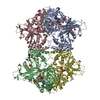
Mass: 785.550 Da / Num. of mol.: 4 / Source method: obtained synthetically / Formula: C27H33N9O15P2 / Comment: FAD*YM
#3: Chemical
ChemComp-SO4 / SULFATE ION / Sulfate


Mass: 96.063 Da / Num. of mol.: 17 / Source method: obtained synthetically / Formula: SO4
#4: Chemical ChemComp-ACT / ACETATE ION / Acetate


Mass: 59.044 Da / Num. of mol.: 1 / Source method: obtained synthetically / Formula: C2H3O2
#5: Chemical ChemComp-GOL / GLYCEROL / GLYCERIN / PROPANE-1,2,3-TRIOL / Glycerol


Mass: 92.094 Da / Num. of mol.: 1 / Source method: obtained synthetically / Formula: C3H8O3
#6: Water ChemComp-HOH / water / Water


Mass: 18.015 Da / Num. of mol.: 516 / Source method: isolated from a natural source / Formula: H2O

-
Details

Sequence detailsAUTHOR STATES THAT A429T IS AN UNINTENDED MUTATION ON THE SURFACE OF THE PROTEIN.

-
Experimental details

-
Experiment

ExperimentMethod: X-RAY DIFFRACTION / Number of used crystals: 1

-
Sample preparation

CrystalDensity Matthews: 4.79 Å3/Da / Density % sol: 74.33 %
Crystal growTemperature: 298 K / Method: vapor diffusion, sitting drop / pH: 4.5
Details: 1.5M ammonium sulfate, 0.1M sodium acetate, 5mM L-cysteine, 0.5mM THP , pH 4.5, VAPOR DIFFUSION, SITTING DROP, temperature 298.0K

-
Data collection

DiffractionMean temperature: 100 K
Diffraction sourceSource: SYNCHROTRON / Site: APS / Beamline: 19-ID / Wavelength: 0.97915 Å
DetectorType: ADSC QUANTUM 315 / Detector: CCD / Date: Aug 6, 2011 / Details: Rosenbaum-Rock vertical focusing mirror
RadiationMonochromator: Rosenbaum-Rock high-resolution double-crystal monochromator
Protocol: SINGLE WAVELENGTH / Monochromatic (M) / Laue (L): M / Scattering type: x-ray
Radiation wavelengthWavelength: 0.97915 Å / Relative weight: 1
ReflectionResolution: 2.35→50 Å / Num. obs: 167455 / % possible obs: 90.5 % / Redundancy: 5.4 % / Rmerge(I) obs: 0.086 / Χ2: 1.017 / Net I/σ(I): 8.4
Reflection shell
Resolution (Å)Redundancy (%)Rmerge(I) obsNum. unique allΧ2Diffraction-ID% possible all
2.35-2.435.40.568166670.59191.1
2.43-2.535.40.427166640.593191.3
2.53-2.655.40.325166950.615191.3
2.65-2.795.40.24167850.644191.5
2.79-2.965.40.163167700.674191.3
2.96-3.195.40.111167990.811191.3
3.19-3.515.40.08167601.155190.7
3.51-4.025.40.069166941.884190
4.02-5.065.30.054166592.057189
5.06-505.40.034169621.166187.3

-
Processing

Software
NameVersionClassificationNB
SCALEPACKdata scaling
PHENIX1.6.2_432refinement
PDB_EXTRACT3.1data extraction
ADSCQuantumdata collection
HKL-3000data reduction
PHENIXphasing
RefinementMethod to determine structure: SAD / Resolution: 2.35→49.421 Å / Occupancy max: 1 / Occupancy min: 0 / FOM work R set: 0.8482 / SU ML: 0.33 / σ(F): 0 / Phase error: 21.91 / Stereochemistry target values: ML
RfactorNum. reflection% reflectionSelection details
Rfree0.2183 8395 5.02 %random
Rwork0.1881 ---
obs0.1896 167290 90.37 %-
Solvent computationShrinkage radii: 0.95 Å / VDW probe radii: 1.2 Å / Solvent model: FLAT BULK SOLVENT MODEL / Bsol: 31.152 Å2 / ksol: 0.354 e/Å3
Displacement parametersBiso max: 144.47 Å2 / Biso mean: 34.984 Å2 / Biso min: 7.15 Å2
Baniso -1Baniso -2Baniso -3
1--5.6926 Å20 Å20 Å2
2---5.6926 Å2-0 Å2
3---11.3853 Å2
Refinement stepCycle: LAST / Resolution: 2.35→49.421 Å
ProteinNucleic acidLigandSolventTotal
Num. atoms15569 0 307 516 16392
Refine LS restraints
Refine-IDTypeDev idealNumber
X-RAY DIFFRACTIONf_bond_d0.00716302
X-RAY DIFFRACTIONf_angle_d1.06422277
X-RAY DIFFRACTIONf_chiral_restr0.0682435
X-RAY DIFFRACTIONf_plane_restr0.0052870
X-RAY DIFFRACTIONf_dihedral_angle_d14.8375969
LS refinement shell

Refine-ID: X-RAY DIFFRACTION / Total num. of bins used: 10

Resolution (Å)Rfactor RfreeNum. reflection RfreeRfactor RworkNum. reflection RworkNum. reflection all% reflection obs (%)
2.35-2.43070.29748050.2486156961650190
2.4307-2.5280.28018410.2198158361667791
2.528-2.6430.25158620.2079158381670091
2.643-2.78240.24198190.2015159751679491
2.7824-2.95670.24527990.2054159611676091
2.9567-3.18490.25198660.2146159411680791
3.1849-3.50540.2418670.2126158901675791
3.5054-4.01240.20927980.1791158831668190
4.0124-5.05440.16178610.1374158191668089
5.0544-49.43220.18958770.1769160561693387
Refinement TLS params.

Method: refined / Refine-ID: X-RAY DIFFRACTION

IDL112)L122)L132)L222)L232)L332)S11 (Å °)S12 (Å °)S13 (Å °)S21 (Å °)S22 (Å °)S23 (Å °)S31 (Å °)S32 (Å °)S33 (Å °)T112)T122)T132)T222)T232)T332)Origin x (Å)Origin y (Å)Origin z (Å)
10.67660.0016-0.2690.0759-0.0280.23610.02120.1918-0.049-0.0033-0.0226-0.0769-0.0211-0.02940.00390.05750.04730.02510.1926-0.0290.122471.423577.7283159.3665
20.2792-0.25860.01210.4220.01980.2317-0.0788-0.0460.04790.09870.04130.062-0.0023-0.0020.00820.08580.04770.02230.0724-0.02230.100624.4064103.2378211.9273
30.87210.1761-0.08520.15430.06940.324-0.03260.3585-0.1832-0.0010.06990.09040.1009-0.0236-0.0618-0.03880.0339-0.07170.2199-0.10250.055925.5668.0432149.5188
40.3171-0.25460.02970.68670.06590.1972-0.0934-0.0622-0.12560.22940.11710.1280.0234-0.0074-0.00670.17090.08630.05310.11840.07910.131241.926859.7539222.1384
Refinement TLS group
IDRefine-IDRefine TLS-IDSelection detailsAuth asym-IDAuth seq-ID
1X-RAY DIFFRACTION1chain AA3 - 507
2X-RAY DIFFRACTION2chain BB3 - 507
3X-RAY DIFFRACTION3chain CC3 - 506
4X-RAY DIFFRACTION4chain DD3 - 506

+
About Yorodumi

-
News

-
Feb 9, 2022. New format data for meta-information of EMDB entries

New format data for meta-information of EMDB entries

  • Version 3 of the EMDB header file is now the official format.
  • The previous official version 1.9 will be removed from the archive.

Related info.:EMDB header

External links:wwPDB to switch to version 3 of the EMDB data model

-
Aug 12, 2020. Covid-19 info

Covid-19 info

URL: https://pdbj.org/emnavi/covid19.php

New page: Covid-19 featured information page in EM Navigator.

Related info.:Covid-19 info / Mar 5, 2020. Novel coronavirus structure data

+
Mar 5, 2020. Novel coronavirus structure data

Novel coronavirus structure data

Related info.:Yorodumi Speices / Aug 12, 2020. Covid-19 info

External links:COVID-19 featured content - PDBj / Molecule of the Month (242):Coronavirus Proteases

+
Jan 31, 2019. EMDB accession codes are about to change! (news from PDBe EMDB page)

EMDB accession codes are about to change! (news from PDBe EMDB page)

  • The allocation of 4 digits for EMDB accession codes will soon come to an end. Whilst these codes will remain in use, new EMDB accession codes will include an additional digit and will expand incrementally as the available range of codes is exhausted. The current 4-digit format prefixed with “EMD-” (i.e. EMD-XXXX) will advance to a 5-digit format (i.e. EMD-XXXXX), and so on. It is currently estimated that the 4-digit codes will be depleted around Spring 2019, at which point the 5-digit format will come into force.
  • The EM Navigator/Yorodumi systems omit the EMD- prefix.

Related info.:Q: What is EMD? / ID/Accession-code notation in Yorodumi/EM Navigator

External links:EMDB Accession Codes are Changing Soon! / Contact to PDBj

+
Jul 12, 2017. Major update of PDB

Major update of PDB

  • wwPDB released updated PDB data conforming to the new PDBx/mmCIF dictionary.
  • This is a major update changing the version number from 4 to 5, and with Remediation, in which all the entries are updated.
  • In this update, many items about electron microscopy experimental information are reorganized (e.g. em_software).
  • Now, EM Navigator and Yorodumi are based on the updated data.

External links:wwPDB Remediation / Enriched Model Files Conforming to OneDep Data Standards Now Available in the PDB FTP Archive

-
Yorodumi

Thousand views of thousand structures

  • Yorodumi is a browser for structure data from EMDB, PDB, SASBDB, etc.
  • This page is also the successor to EM Navigator detail page, and also detail information page/front-end page for Omokage search.
  • The word "yorodu" (or yorozu) is an old Japanese word meaning "ten thousand". "mi" (miru) is to see.

Related info.:EMDB / PDB / SASBDB / Comparison of 3 databanks / Yorodumi Search / Aug 31, 2016. New EM Navigator & Yorodumi / Yorodumi Papers / Jmol/JSmol / Function and homology information / Changes in new EM Navigator and Yorodumi

Read more