 Movie
Movie Controller

[English] 日本語
 Yorodumi
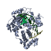
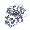
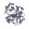
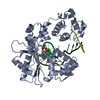
- PDB-3uq2: Crystal structure of the post-catalytic product complex of polyme... -
+ Open data

- Basic information
| Entry | Database: PDB / ID: 3uq2 | ||||||
|---|---|---|---|---|---|---|---|
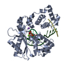
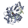
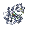
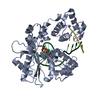
| Title | Crystal structure of the post-catalytic product complex of polymerase lambda with an rCMP inserted opposite a templating G and dAMP inserted opposite a templating T at the primer terminus. | ||||||
 Components Components |
| ||||||
 Keywords Keywords | TRANSFERASE / LYASE/DNA / DNA polymerase lambda / ribonucleotide incorporation / Protein Conformation / LYASE-DNA complex | ||||||
| Function / homology |  Function and homology information Function and homology informationDNA biosynthetic process / Lyases; Carbon-oxygen lyases; Other carbon-oxygen lyases / 5'-deoxyribose-5-phosphate lyase activity / somatic hypermutation of immunoglobulin genes / base-excision repair, gap-filling / nucleotide-excision repair / Nonhomologous End-Joining (NHEJ) / double-strand break repair via homologous recombination / double-strand break repair via nonhomologous end joining / site of double-strand break ...DNA biosynthetic process / Lyases; Carbon-oxygen lyases; Other carbon-oxygen lyases / 5'-deoxyribose-5-phosphate lyase activity / somatic hypermutation of immunoglobulin genes / base-excision repair, gap-filling / nucleotide-excision repair / Nonhomologous End-Joining (NHEJ) / double-strand break repair via homologous recombination / double-strand break repair via nonhomologous end joining / site of double-strand break / DNA-directed DNA polymerase / DNA-directed DNA polymerase activity / DNA replication / DNA binding / nucleoplasm / metal ion binding / nucleus Similarity search - Function | ||||||
| Biological species |  Homo sapiens (human) Homo sapiens (human) | ||||||
| Method |  X-RAY DIFFRACTION / SYNCHROTRON / FOURIER SYNTHESIS / Resolution: 2.25 Å X-RAY DIFFRACTION / SYNCHROTRON / FOURIER SYNTHESIS / Resolution: 2.25 Å | ||||||
 Authors Authors | Gosavi, R.A. / Moon, A.F. / Kunkel, T.A. / Pedersen, L.C. / Bebenek, K. | ||||||
 Citation Citation | Journal: Nucleic Acids Res. / Year: 2012 Title: The catalytic cycle for ribonucleotide incorporation by human DNA Pol lambda Authors: Gosavi, R.A. / Moon, A.F. / Kunkel, T.A. / Pedersen, L.C. / Bebenek, K. | ||||||
| History |
|
- Structure visualization
| Structure viewer | Molecule: MolmilJmol/JSmol |
|---|
- Downloads & links
-Download
| PDBx/mmCIF format | 3uq2.cif.gz | 95.8 KB | Display | PDBx/mmCIF format |
|---|---|---|---|---|
| PDB format | pdb3uq2.ent.gz | 66.7 KB | Display | PDB format |
| PDBx/mmJSON format | 3uq2.json.gz | Tree view | PDBx/mmJSON format | |
| Others |  Other downloads Other downloads |
-Validation report
| Arichive directory | https://data.pdbj.org/pub/pdb/validation_reports/uq/3uq2ftp://data.pdbj.org/pub/pdb/validation_reports/uq/3uq2 | HTTPS FTP |
|---|
-Related structure data
| Related structure data | 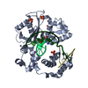 3upqC 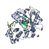 3uq0C 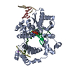 4fo6C  3mgiS S: Starting model for refinement C: citing same article ( |
|---|---|
| Similar structure data |


-Links
 PDBj
PDBj


- Assembly
| Deposited unit | 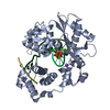
| ||||||||
|---|---|---|---|---|---|---|---|---|---|
| 1 |
| ||||||||
| Unit cell |
|
-Components
-DNA chain , 2 types, 2 molecules TD
| #2: DNA chain | Mass: 3365.196 Da / Num. of mol.: 1 / Source method: obtained synthetically |
|---|---|
| #4: DNA chain | Mass: 1191.818 Da / Num. of mol.: 1 / Source method: obtained synthetically |
-Protein / DNA/RNA hybrid , 2 types, 2 molecules AP
| #1: Protein | Mass: 36703.977 Da / Num. of mol.: 1 / Fragment: Loop mutant of DNA polymerase lambda / Mutation: SQEENGQQQ to KGET Source method: isolated from a genetically manipulated source Source: (gene. exp.) Homo sapiens (human) / Gene: POLL / Production host:  References: UniProt: Q9UGP5, DNA-directed DNA polymerase, Lyases; Carbon-oxygen lyases; Other carbon-oxygen lyases |
|---|---|
| #3: DNA/RNA hybrid | Mass: 2122.425 Da / Num. of mol.: 1 / Source method: obtained synthetically |
-Non-polymers , 4 types, 116 molecules 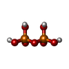


| #5: Chemical | ChemComp-PPV /  Mass: 177.975 Da / Num. of mol.: 1 / Source method: obtained synthetically / Formula: H4O7P2 Mass: 177.975 Da / Num. of mol.: 1 / Source method: obtained synthetically / Formula: H4O7P2 |
|---|---|
| #6: Chemical | ChemComp-MG /  Mass: 24.305 Da / Num. of mol.: 1 / Source method: obtained synthetically / Formula: Mg Mass: 24.305 Da / Num. of mol.: 1 / Source method: obtained synthetically / Formula: Mg |
| #7: Chemical | ChemComp-NA /  Mass: 22.990 Da / Num. of mol.: 1 / Source method: obtained synthetically / Formula: Na Mass: 22.990 Da / Num. of mol.: 1 / Source method: obtained synthetically / Formula: Na |
| #8: Water | ChemComp-HOH / Mass: 18.015 Da / Num. of mol.: 113 / Source method: isolated from a natural source / Formula: H2O |
-Details
| Sequence details | AUTHORS STATE THAT THIS IS A DELETION-SUBSTITUTION MUTANT, WITH RESIDUES SQEENGQQQ IN THE WILDTYPE ...AUTHORS STATE THAT THIS IS A DELETION-SUBSTITUTI |
|---|
-Experimental details
-Experiment
| Experiment | Method: X-RAY DIFFRACTION / Number of used crystals: 1 |
|---|
- Sample preparation
| Crystal | Density Matthews: 2.79 Å3/Da / Density % sol: 55.95 % |
|---|---|
| Crystal grow | Temperature: 293 K / Method: vapor diffusion, sitting drop / pH: 5.6 Details: 170mM ammonium acetate, 85mM sodium citrate tribasic dihydrate pH 5.6, 25.5% (w/v) PEG4000, 15% (v/v) glycerol, VAPOR DIFFUSION, SITTING DROP, temperature 293K |
-Data collection
| Diffraction | Mean temperature: 100 K |
|---|---|
| Diffraction source | Source: SYNCHROTRON / Site: APS  / Beamline: 22-BM / Wavelength: 1 Å / Beamline: 22-BM / Wavelength: 1 Å |
| Detector | Type: MARMOSAIC 225 mm CCD / Detector: CCD / Date: Apr 24, 2011 |
| Radiation | Monochromator: GRAPHITE / Protocol: SINGLE WAVELENGTH / Monochromatic (M) / Laue (L): M / Scattering type: x-ray |
| Radiation wavelength | Wavelength: 1 Å / Relative weight: 1 |
| Reflection | Resolution: 2.25→50 Å / Num. obs: 23003 / % possible obs: 93.1 % / Observed criterion σ(F): 0 / Observed criterion σ(I): 0 / Redundancy: 6.2 % / Biso Wilson estimate: 41.68 Å2 / Rsym value: 0.069 / Net I/σ(I): 23.7 |
| Reflection shell | Resolution: 2.25→2.29 Å / Redundancy: 5.5 % / Mean I/σ(I) obs: 5.3 / Rsym value: 0.288 / % possible all: 69.7 |
- Processing
| Software |
| |||||||||||||||||||||||||||||||||||||||||||||||||||||||||||||||
|---|---|---|---|---|---|---|---|---|---|---|---|---|---|---|---|---|---|---|---|---|---|---|---|---|---|---|---|---|---|---|---|---|---|---|---|---|---|---|---|---|---|---|---|---|---|---|---|---|---|---|---|---|---|---|---|---|---|---|---|---|---|---|---|---|
| Refinement | Method to determine structure: FOURIER SYNTHESIS Starting model: 3MGI Resolution: 2.25→27.13 Å / SU ML: 0.56 / Cross valid method: THROUGHOUT / σ(F): 1.34 / Phase error: 30.64 / Stereochemistry target values: ML
| |||||||||||||||||||||||||||||||||||||||||||||||||||||||||||||||
| Solvent computation | Shrinkage radii: 0.86 Å / VDW probe radii: 1.1 Å / Solvent model: FLAT BULK SOLVENT MODEL / Bsol: 59.528 Å2 / ksol: 0.356 e/Å3 | |||||||||||||||||||||||||||||||||||||||||||||||||||||||||||||||
| Displacement parameters |
| |||||||||||||||||||||||||||||||||||||||||||||||||||||||||||||||
| Refinement step | Cycle: LAST / Resolution: 2.25→27.13 Å
| |||||||||||||||||||||||||||||||||||||||||||||||||||||||||||||||
| Refine LS restraints |
| |||||||||||||||||||||||||||||||||||||||||||||||||||||||||||||||
| LS refinement shell |
|
