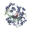
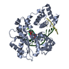
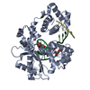
DNA biosynthetic process / Lyases; Carbon-oxygen lyases; Other carbon-oxygen lyases / 5'-deoxyribose-5-phosphate lyase activity / somatic hypermutation of immunoglobulin genes / base-excision repair, gap-filling / nucleotide-excision repair / Nonhomologous End-Joining (NHEJ) / double-strand break repair via homologous recombination / double-strand break repair via nonhomologous end joining / site of double-strand break ...DNA biosynthetic process / Lyases; Carbon-oxygen lyases; Other carbon-oxygen lyases / 5'-deoxyribose-5-phosphate lyase activity / somatic hypermutation of immunoglobulin genes / base-excision repair, gap-filling / nucleotide-excision repair / Nonhomologous End-Joining (NHEJ) / double-strand break repair via homologous recombination / double-strand break repair via nonhomologous end joining / site of double-strand break / DNA-directed DNA polymerase / DNA-directed DNA polymerase activity / DNA replication / DNA binding / nucleoplasm / metal ion binding / nucleus Similarity search - Function
Beta Polymerase; domain 3 / DNA polymerase, thumb domain / DNA polymerase family X, beta-like / DNA polymerase beta, N-terminal domain-like / DNA polymerase beta, palm domain / DNA polymerase beta palm / DNA polymerase lambda, fingers domain / Fingers domain of DNA polymerase lambda / DNA-directed DNA polymerase X / DNA polymerase X family ...Beta Polymerase; domain 3 / DNA polymerase, thumb domain / DNA polymerase family X, beta-like / DNA polymerase beta, N-terminal domain-like / DNA polymerase beta, palm domain / DNA polymerase beta palm / DNA polymerase lambda, fingers domain / Fingers domain of DNA polymerase lambda / DNA-directed DNA polymerase X / DNA polymerase X family / DNA polymerase family X, binding site / DNA polymerase family X signature. / DNA polymerase beta-like, N-terminal domain / DNA polymerase lambda lyase domain superfamily / Helix-hairpin-helix domain / DNA polymerase family X / DNA polymerase beta, thumb domain / DNA polymerase, thumb domain superfamily / DNA polymerase beta thumb / Beta Polymerase, domain 2 / Beta Polymerase; domain 2 / BRCT domain profile. / BRCT domain / BRCT domain superfamily / Nucleotidyltransferase superfamily / 5' to 3' exonuclease, C-terminal subdomain / DNA polymerase; domain 1 / 2-Layer Sandwich / Orthogonal Bundle / Mainly Alpha / Alpha Beta Similarity search - Domain/homology
In the structure databanks used in Yorodumi, some data are registered as the other names, "COVID-19 virus" and "2019-nCoV". Here are the details of the virus and the list of structure data.
Jan 31, 2019. EMDB accession codes are about to change! (news from PDBe EMDB page)
EMDB accession codes are about to change! (news from PDBe EMDB page)
The allocation of 4 digits for EMDB accession codes will soon come to an end. Whilst these codes will remain in use, new EMDB accession codes will include an additional digit and will expand incrementally as the available range of codes is exhausted. The current 4-digit format prefixed with “EMD-” (i.e. EMD-XXXX) will advance to a 5-digit format (i.e. EMD-XXXXX), and so on. It is currently estimated that the 4-digit codes will be depleted around Spring 2019, at which point the 5-digit format will come into force.
The EM Navigator/Yorodumi systems omit the EMD- prefix.
Related info.:Q: What is EMD? / ID/Accession-code notation in Yorodumi/EM Navigator
Yorodumi is a browser for structure data from EMDB, PDB, SASBDB, etc.
This page is also the successor to EM Navigator detail page, and also detail information page/front-end page for Omokage search.
The word "yorodu" (or yorozu) is an old Japanese word meaning "ten thousand". "mi" (miru) is to see.
Related info.:EMDB / PDB / SASBDB / Comparison of 3 databanks / Yorodumi Search / Aug 31, 2016. New EM Navigator & Yorodumi / Yorodumi Papers / Jmol/JSmol / Function and homology information / Changes in new EM Navigator and Yorodumi
 Movie
Movie Controller

 Yorodumi
 Open data
 Basic information
 Components
 Keywords
 Function and homology information
 Homo sapiens (human)
 X-RAY DIFFRACTION /
  Authors
 Citation
 Structure visualization
 Downloads & links
 Other downloads
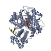


 PDBj
PDBj


 Assembly


 Mass: 505.208 Da / Num. of mol.: 1 / Source method: obtained synthetically / Formula: C11H18N5O12P3
 Mass: 24.305 Da / Num. of mol.: 1 / Source method: obtained synthetically / Formula: Mg
 Mass: 22.990 Da / Num. of mol.: 3 / Source method: obtained synthetically / Formula: Na
 Mass: 122.143 Da / Num. of mol.: 1 / Source method: obtained synthetically / Formula: C4H12NO3 / Comment: pH buffer*YM
 Sample preparation
 Processing