 Movie
Movie Controller

[English] 日本語
 Yorodumi
- PDB-3ulr: Lysozyme contamination facilitates crystallization of a hetero-tr... -
+ Open data

- Basic information
| Entry | Database: PDB / ID: 3ulr | ||||||
|---|---|---|---|---|---|---|---|
| Title | Lysozyme contamination facilitates crystallization of a hetero-trimericCortactin:Arg:Lysozyme complex | ||||||
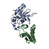
 Components Components |
| ||||||
 Keywords Keywords | HYDROLASE / PROTEIN BINDING / SH3 / Protein-protein interaction | ||||||
| Function / homology |  Function and homology information Function and homology informationlamellipodium organization / site of polarized growth / Arp2/3 complex binding / mitotic spindle midzone / regulation of cell projection assembly / Role of ABL in ROBO-SLIT signaling / modification of postsynaptic actin cytoskeleton / regulation of mitophagy / profilin binding / modification of postsynaptic structure ...lamellipodium organization / site of polarized growth / Arp2/3 complex binding / mitotic spindle midzone / regulation of cell projection assembly / Role of ABL in ROBO-SLIT signaling / modification of postsynaptic actin cytoskeleton / regulation of mitophagy / profilin binding / modification of postsynaptic structure / regulation of actin filament polymerization / positive regulation of smooth muscle contraction / substrate-dependent cell migration, cell extension / regulation of cell motility / focal adhesion assembly / positive regulation of chemotaxis / positive regulation of establishment of T cell polarity / proline-rich region binding / postsynaptic actin cytoskeleton / podosome / negative regulation of Rho protein signal transduction / dendritic spine maintenance / regulation of axon extension / cortical actin cytoskeleton / positive regulation of actin filament polymerization / cortical cytoskeleton / exploration behavior / regulation of endocytosis / actin monomer binding / RAC3 GTPase cycle / positive regulation of T cell migration / regulation of cell adhesion / cellular response to retinoic acid / ruffle / phosphotyrosine residue binding / peptidyl-tyrosine phosphorylation / neuron projection morphogenesis / RAC1 GTPase cycle / clathrin-coated pit / voltage-gated potassium channel complex / Lactose synthesis / Antimicrobial peptides / Negative regulation of FLT3 / receptor-mediated endocytosis / Neutrophil degranulation / beta-N-acetylglucosaminidase activity / protein modification process / negative regulation of extrinsic apoptotic signaling pathway / regulation of autophagy / regulation of actin cytoskeleton organization / non-specific protein-tyrosine kinase / actin filament / cell motility / non-membrane spanning protein tyrosine kinase activity / intracellular protein transport / positive regulation of neuron projection development / enzyme activator activity / epidermal growth factor receptor signaling pathway / cell wall macromolecule catabolic process / lysozyme / lysozyme activity / actin filament binding / cell junction / manganese ion binding / cell migration / lamellipodium / actin cytoskeleton / growth cone / positive regulation of cytosolic calcium ion concentration / actin cytoskeleton organization / protein tyrosine kinase activity / cellular response to oxidative stress / cell cortex / killing of cells of another organism / defense response to Gram-negative bacterium / dendritic spine / phospholipase C-activating G protein-coupled receptor signaling pathway / protein kinase activity / cell adhesion / defense response to bacterium / postsynapse / defense response to Gram-positive bacterium / focal adhesion / glutamatergic synapse / magnesium ion binding / enzyme binding / endoplasmic reticulum / Golgi apparatus / signal transduction / : / ATP binding / identical protein binding / plasma membrane / cytoplasm / cytosol Similarity search - Function | ||||||
| Biological species |    Homo sapiens (human) Homo sapiens (human) | ||||||
| Method |  X-RAY DIFFRACTION / SYNCHROTRON / MOLECULAR REPLACEMENT / Resolution: 1.65 Å X-RAY DIFFRACTION / SYNCHROTRON / MOLECULAR REPLACEMENT / Resolution: 1.65 Å | ||||||
 Authors Authors | Liu, W. / MacGrath, S. / Koleske, A.J. / Boggon, T.J. | ||||||
 Citation Citation | Journal: Acta Crystallogr.,Sect.F / Year: 2012 Title: Lysozyme contamination facilitates crystallization of a heterotrimeric cortactin-Arg-lysozyme complex. Authors: Liu, W. / Macgrath, S.M. / Koleske, A.J. / Boggon, T.J. | ||||||
| History |
|
- Structure visualization
| Structure viewer | Molecule: MolmilJmol/JSmol |
|---|
- Downloads & links
-Download
| PDBx/mmCIF format | 3ulr.cif.gz | 59.9 KB | Display | PDBx/mmCIF format |
|---|---|---|---|---|
| PDB format | pdb3ulr.ent.gz | 43.2 KB | Display | PDB format |
| PDBx/mmJSON format | 3ulr.json.gz | Tree view | PDBx/mmJSON format | |
| Others |  Other downloads Other downloads |
-Validation report
| Arichive directory | https://data.pdbj.org/pub/pdb/validation_reports/ul/3ulrftp://data.pdbj.org/pub/pdb/validation_reports/ul/3ulr | HTTPS FTP |
|---|
-Related structure data


-Links
 PDBj
PDBj


- Assembly
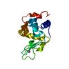
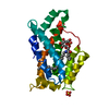
| Deposited unit | 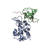
| ||||||||
|---|---|---|---|---|---|---|---|---|---|
| 1 |
| ||||||||
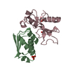
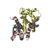
| Unit cell |
|
-Components
| #1: Protein | Mass: 14331.160 Da / Num. of mol.: 1 Source method: isolated from a genetically manipulated source Details: American Bioanalytical AB01178 / Source: (gene. exp.) |
|---|---|
| #2: Protein | Mass: 7202.915 Da / Num. of mol.: 1 / Fragment: SH3 domain (UNP residues 487-546) Source method: isolated from a genetically manipulated source Source: (gene. exp.)  |
| #3: Protein/peptide | Mass: 1869.252 Da / Num. of mol.: 1 / Fragment: PXXP1 (UNP residues 563-579) / Source method: obtained synthetically / Source: (synth.) Homo sapiens (human) / References: UniProt: P42684 |
| #4: Water | ChemComp-HOH /  Mass: 18.015 Da / Num. of mol.: 292 / Source method: isolated from a natural source / Formula: H2O Mass: 18.015 Da / Num. of mol.: 292 / Source method: isolated from a natural source / Formula: H2O |
| Has protein modification | Y |
-Experimental details
-Experiment
| Experiment | Method: X-RAY DIFFRACTION / Number of used crystals: 1 |
|---|
- Sample preparation
| Crystal | Density Matthews: 2.28 Å3/Da / Density % sol: 46.08 % |
|---|---|
| Crystal grow | Temperature: 297 K / Method: vapor diffusion, hanging drop / pH: 6 Details: 1.0M Na Citrate, pH 6.0, VAPOR DIFFUSION, HANGING DROP, temperature 297K |
-Data collection
| Diffraction | Mean temperature: 100 K |
|---|---|
| Diffraction source | Source: SYNCHROTRON / Site: NSLS  / Beamline: X6A / Wavelength: 1.0781 Å / Beamline: X6A / Wavelength: 1.0781 Å |
| Detector | Type: ADSC QUANTUM 270 / Detector: CCD / Date: Oct 5, 2010 Details: Si(111) channel cut monochromator Toroidal focusing mirror |
| Radiation | Monochromator: Si 111 / Protocol: SINGLE WAVELENGTH / Monochromatic (M) / Laue (L): M / Scattering type: x-ray |
| Radiation wavelength | Wavelength: 1.0781 Å / Relative weight: 1 |
| Reflection | Resolution: 1.65→20 Å / Num. all: 25122 / Num. obs: 23866 / % possible obs: 95 % / Observed criterion σ(F): 0 / Observed criterion σ(I): 0 / Redundancy: 5.7 % / Biso Wilson estimate: 20.7 Å2 / Rsym value: 0.087 / Net I/σ(I): 14.9 |
| Reflection shell | Resolution: 1.65→1.71 Å / Redundancy: 2.9 % / Mean I/σ(I) obs: 2.5 / Num. unique all: 1704 / Rsym value: 0.35 / % possible all: 65.8 |
- Processing
| Software |
| |||||||||||||||||||||||||||||||||||||||||||||||||||||||||||||||||
|---|---|---|---|---|---|---|---|---|---|---|---|---|---|---|---|---|---|---|---|---|---|---|---|---|---|---|---|---|---|---|---|---|---|---|---|---|---|---|---|---|---|---|---|---|---|---|---|---|---|---|---|---|---|---|---|---|---|---|---|---|---|---|---|---|---|---|
| Refinement | Method to determine structure: MOLECULAR REPLACEMENT Starting model: 2D1X and 3M3U Resolution: 1.65→20 Å / Cor.coef. Fo:Fc: 0.964 / Cor.coef. Fo:Fc free: 0.945 / SU B: 2.478 / SU ML: 0.083 / Cross valid method: THROUGHOUT / ESU R Free: 0.11 / Stereochemistry target values: Engh & Huber
| |||||||||||||||||||||||||||||||||||||||||||||||||||||||||||||||||
| Solvent computation | Ion probe radii: 0.8 Å / Shrinkage radii: 0.8 Å / VDW probe radii: 1.4 Å / Solvent model: MASK | |||||||||||||||||||||||||||||||||||||||||||||||||||||||||||||||||
| Displacement parameters | Biso mean: 24.167 Å2
| |||||||||||||||||||||||||||||||||||||||||||||||||||||||||||||||||
| Refinement step | Cycle: LAST / Resolution: 1.65→20 Å
| |||||||||||||||||||||||||||||||||||||||||||||||||||||||||||||||||
| Refine LS restraints |
| |||||||||||||||||||||||||||||||||||||||||||||||||||||||||||||||||
| LS refinement shell | Resolution: 1.651→1.694 Å / Total num. of bins used: 20
|
