Mass: 18.015 Da / Num. of mol.: 87 / Source method: isolated from a natural source / Formula: H2O
Has protein modification
Y
Sequence details
THE CONSTRUCT (RESIDUES 82-244) WAS EXPRESSED WITH AN N-TERMINAL EXPRESSION / PURIFICATION TAG ...THE CONSTRUCT (RESIDUES 82-244) WAS EXPRESSED WITH AN N-TERMINAL EXPRESSION / PURIFICATION TAG MGSDKIHHHHHHAEIGTGFPFDPHYVEVLGERMHYVDVGPRDGTPVLFLHGNPTSSYVWRNIIPHVA PTHRCIAPDLIGMGKSDKPDLGYFFDDHVRFMDAFIEALGLEEVVLVIHDWGSALGFHWAKRNPERV KGIAFMEFIRPIPTWDEWPEFARETFQAFRTTDVGRKLIIDQNVFIEGTLPMGVVRPLTEVEMDHYR EPFLNPVDREPLWRFPNELPIAGEPANIVALVEEYMDWLHQSPVPKLLFWGTPGVLIPPAEAARLAK SLPNCKAVDIGPGLNLLQEDNPDLIGSEIARWLSTLEISGEPTTHENLYFQG. THE TAG WAS REMOVED WITH TEV PROTEASE LEAVING ONLY A GLYCINE (0) FOLLOWED BY THE TARGET SEQUENCE. WHILE THE CLONED SEQUENCE MATCHES ISOFORM 2 (ALSO KNOWN AS T) FOR UNIPROTKB ID P39688, RESIDUE NUMBERING IS BASED ON THE UNIPROT "CANONICAL" ISOFORM 1 (ALSO KNOWN AS B).
-
Experimental details
-
Experiment
Experiment
Method: X-RAY DIFFRACTION / Number of used crystals: 1
-
Sample preparation
Crystal
Density Matthews: 2.89 Å3/Da / Density % sol: 57.4 %
Crystal grow
Temperature: 277 K / Method: vapor diffusion, sitting drop / pH: 7.5 Details: 4.3M NaCl, 0.1M HEPES pH 7.5, NANODROP, VAPOR DIFFUSION, SITTING DROP, temperature 277K
Monochromator: Double Crystal Si(111) / Protocol: SINGLE WAVELENGTH / Monochromatic (M) / Laue (L): M / Scattering type: x-ray
Radiation wavelength
Wavelength: 0.9793 Å / Relative weight: 1
Reflection
Resolution: 1.98→43.876 Å / Num. obs: 14960 / % possible obs: 97.8 % / Observed criterion σ(I): -3 / Biso Wilson estimate: 32.893 Å2 / Rmerge(I) obs: 0.058 / Net I/σ(I): 15.73
Reflection shell
Diffraction-ID: 1
Resolution (Å)
Highest resolution (Å)
Rmerge(I) obs
Mean I/σ(I) obs
Num. measured obs
Num. unique obs
% possible all
1.98-2.05
0.811
1.8
6306
1420
96.9
2.05-2.13
0.535
3.1
6356
1411
95.1
2.13-2.23
0.402
3.8
7506
1545
98.9
2.23-2.35
0.32
5.2
6548
1439
93.4
2.35-2.49
0.208
7
7045
1435
99.9
2.49-2.69
0.144
10.1
7381
1551
98
2.69-2.96
0.081
16.1
7421
1521
99.7
2.96-3.38
0.042
27.2
7152
1495
99.6
3.38-4.25
0.033
37.1
6611
1477
96.9
4.25
0.028
42.5
7413
1616
99.5
-
Phasing
Phasing
Method: molecular replacement
-
Processing
Software
Name
Version
Classification
NB
MolProbity
3beta29
modelbuilding
PDB_EXTRACT
3.1
dataextraction
MOLREP
phasing
XSCALE
December6, 2010
datascaling
BUSTER-TNT
2.10.0
refinement
XDS
datareduction
BUSTER
2.10.0
refinement
Refinement
Method to determine structure: MOLECULAR REPLACEMENT / Resolution: 1.98→43.876 Å / Cor.coef. Fo:Fc: 0.9501 / Cor.coef. Fo:Fc free: 0.9344 / Occupancy max: 1 / Occupancy min: 0.4 / Cross valid method: THROUGHOUT / σ(F): 0 Details: 1. A MET-INHIBITION PROTOCOL WAS USED FOR SELENOMETHIONINE INCORPORATION DURING PROTEIN EXPRESSION. HOWEVER, THE SE-MET SIDE-CHAIN IS DISORDERED. 2. ATOM RECORD CONTAINS SUM OF TLS AND ...Details: 1. A MET-INHIBITION PROTOCOL WAS USED FOR SELENOMETHIONINE INCORPORATION DURING PROTEIN EXPRESSION. HOWEVER, THE SE-MET SIDE-CHAIN IS DISORDERED. 2. ATOM RECORD CONTAINS SUM OF TLS AND RESIDUAL B FACTORS. ANISOU RECORD CONTAINS SUM OF TLS AND RESIDUAL U FACTORS. 3. GLYCEROL, SODIUM AND CHLORIDE MODELED IS PRESENT IN CRYO/CRYSTALLIZATION CONDITIONS.
In the structure databanks used in Yorodumi, some data are registered as the other names, "COVID-19 virus" and "2019-nCoV". Here are the details of the virus and the list of structure data.
Jan 31, 2019. EMDB accession codes are about to change! (news from PDBe EMDB page)
EMDB accession codes are about to change! (news from PDBe EMDB page)
The allocation of 4 digits for EMDB accession codes will soon come to an end. Whilst these codes will remain in use, new EMDB accession codes will include an additional digit and will expand incrementally as the available range of codes is exhausted. The current 4-digit format prefixed with “EMD-” (i.e. EMD-XXXX) will advance to a 5-digit format (i.e. EMD-XXXXX), and so on. It is currently estimated that the 4-digit codes will be depleted around Spring 2019, at which point the 5-digit format will come into force.
The EM Navigator/Yorodumi systems omit the EMD- prefix.
Related info.:Q: What is EMD? / ID/Accession-code notation in Yorodumi/EM Navigator
Yorodumi is a browser for structure data from EMDB, PDB, SASBDB, etc.
This page is also the successor to EM Navigator detail page, and also detail information page/front-end page for Omokage search.
The word "yorodu" (or yorozu) is an old Japanese word meaning "ten thousand". "mi" (miru) is to see.
Related info.:EMDB / PDB / SASBDB / Comparison of 3 databanks / Yorodumi Search / Aug 31, 2016. New EM Navigator & Yorodumi / Yorodumi Papers / Jmol/JSmol / Function and homology information / Changes in new EM Navigator and Yorodumi
 Movie
Movie Controller

 Yorodumi
 Open data
 Basic information
 Components
 Keywords
 Function and homology information
 X-RAY DIFFRACTION /
  Authors
 Citation
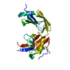
 Structure visualization
 Downloads & links
 Other downloads
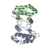


 PDBj
PDBj


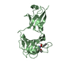
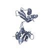
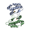
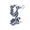
 Assembly
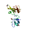


 Mass: 22.990 Da / Num. of mol.: 1 / Source method: obtained synthetically / Formula: Na
 Mass: 35.453 Da / Num. of mol.: 2 / Source method: obtained synthetically / Formula: Cl
 Mass: 92.094 Da / Num. of mol.: 2 / Source method: obtained synthetically / Formula: C3H8O3
 Mass: 18.015 Da / Num. of mol.: 87 / Source method: isolated from a natural source / Formula: H2O
 Sample preparation
 / Beamline: 8.2.2 / Wavelength: 0.9793
  Processing