 Movie
Movie Controller

[English] 日本語
 Yorodumi
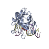
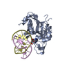
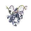
- PDB-3uby: Crystal structure of human alklyadenine DNA glycosylase in a lowe... -
+ Open data

- Basic information
| Entry | Database: PDB / ID: 3uby | ||||||
|---|---|---|---|---|---|---|---|
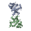
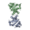
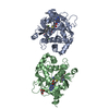
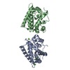
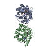
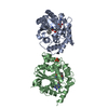
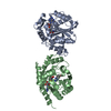
| Title | Crystal structure of human alklyadenine DNA glycosylase in a lower and higher-affinity complex with DNA | ||||||
 Components Components |
| ||||||
 Keywords Keywords | HYDROLASE/DNA / alkyladenine DNA glycosylase fold / AAG / DNA repair / DNA binding / Nucleus / HYDROLASE-DNA complex | ||||||
| Function / homology |  Function and homology information Function and homology informationalkylbase DNA N-glycosylase activity / DNA-3-methyladenine glycosylase II / depurination / DNA N-glycosylase activity / Displacement of DNA glycosylase by APEX1 / DNA alkylation repair / mitochondrial nucleoid / Recognition and association of DNA glycosylase with site containing an affected purine / Cleavage of the damaged purine / base-excision repair ...alkylbase DNA N-glycosylase activity / DNA-3-methyladenine glycosylase II / depurination / DNA N-glycosylase activity / Displacement of DNA glycosylase by APEX1 / DNA alkylation repair / mitochondrial nucleoid / Recognition and association of DNA glycosylase with site containing an affected purine / Cleavage of the damaged purine / base-excision repair / damaged DNA binding / mitochondrion / DNA binding / nucleoplasm / cytosol Similarity search - Function | ||||||
| Biological species |  Homo sapiens (human) Homo sapiens (human) | ||||||
| Method |  X-RAY DIFFRACTION / SYNCHROTRON / MOLECULAR REPLACEMENT / Resolution: 2 Å X-RAY DIFFRACTION / SYNCHROTRON / MOLECULAR REPLACEMENT / Resolution: 2 Å | ||||||
 Authors Authors | Setser, J.W. / Lingaraju, G.M. / Davis, C.A. / Samson, L.D. / Drennan, C.L. | ||||||
 Citation Citation | Journal: Biochemistry / Year: 2012 Title: Searching for DNA lesions: structural evidence for lower- and higher-affinity DNA binding conformations of human alkyladenine DNA glycosylase. Authors: Setser, J.W. / Lingaraju, G.M. / Davis, C.A. / Samson, L.D. / Drennan, C.L. | ||||||
| History |
|
- Structure visualization
| Structure viewer | Molecule: MolmilJmol/JSmol |
|---|
- Downloads & links
-Download
| PDBx/mmCIF format | 3uby.cif.gz | 188.3 KB | Display | PDBx/mmCIF format |
|---|---|---|---|---|
| PDB format | pdb3uby.ent.gz | 145.9 KB | Display | PDB format |
| PDBx/mmJSON format | 3uby.json.gz | Tree view | PDBx/mmJSON format | |
| Others |  Other downloads Other downloads |
-Validation report
| Summary document | 3uby_validation.pdf.gz | 450.7 KB | Display | wwPDB validaton report |
|---|---|---|---|---|
| Full document | 3uby_full_validation.pdf.gz | 454.7 KB | Display | |
| Data in XML | 3uby_validation.xml.gz | 19.5 KB | Display | |
| Data in CIF | 3uby_validation.cif.gz | 27.9 KB | Display | |
| Arichive directory | https://data.pdbj.org/pub/pdb/validation_reports/ub/3ubyftp://data.pdbj.org/pub/pdb/validation_reports/ub/3uby | HTTPS FTP |
-Related structure data
| Related structure data | 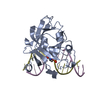 1bnkS S: Starting model for refinement |
|---|---|
| Similar structure data |


-Links
 PDBj
PDBj


- Assembly
| Deposited unit | 
| ||||||||
|---|---|---|---|---|---|---|---|---|---|
| 1 | 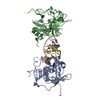
| ||||||||
| Unit cell |
|
-Components
| #1: Protein | Mass: 24314.891 Da / Num. of mol.: 2 / Fragment: DELTA79AAG Source method: isolated from a genetically manipulated source Source: (gene. exp.) Homo sapiens (human) / Gene: MPG, AAG, ANPG, MID1 / Production host:  References: UniProt: P29372, DNA-3-methyladenine glycosylase II #2: DNA chain | Mass: 3966.594 Da / Num. of mol.: 2 / Source method: obtained synthetically #3: Water | ChemComp-HOH / |  Mass: 18.015 Da / Num. of mol.: 250 / Source method: isolated from a natural source / Formula: H2O Mass: 18.015 Da / Num. of mol.: 250 / Source method: isolated from a natural source / Formula: H2O |
|---|
-Experimental details
-Experiment
| Experiment | Method: X-RAY DIFFRACTION / Number of used crystals: 1 |
|---|
- Sample preparation
| Crystal | Density Matthews: 1.97 Å3/Da / Density % sol: 37.46 % |
|---|---|
| Crystal grow | Temperature: 298 K / Method: vapor diffusion, hanging drop Details: An equimolar ratio of delta79AAG and 13-mer single-stranded (ss) EDC DNA were mixed to form a protein-DNA complex concentration of 0.3 mM in the complex buffer (20 mM HEPES-NaOH, pH 7.5, 100 ...Details: An equimolar ratio of delta79AAG and 13-mer single-stranded (ss) EDC DNA were mixed to form a protein-DNA complex concentration of 0.3 mM in the complex buffer (20 mM HEPES-NaOH, pH 7.5, 100 mM NaCl, 0.1 mM EDTA, 5% v/v glycerol and 1 mM DTT). The complex was incubated on ice for 15 min and used for crystallization. Crystals were obtained upon mixing 1 uL of protein-DNA complex and 1 uL of reservoir solution (100 mM BIS-TRIS, pH 5.5, 200 mM cesium chloride and 20% polyethylene glycol (PEG) 3350) over 0.5 ml of reservoir solution. Crystals appeared after incubation for 14 days at 22 degrees C, VAPOR DIFFUSION, HANGING DROP, temperature 298K |
-Data collection
| Diffraction | Mean temperature: 100 K |
|---|---|
| Diffraction source | Source: SYNCHROTRON / Site: ALS  / Beamline: 12.3.1 / Wavelength: 1.116 Å / Beamline: 12.3.1 / Wavelength: 1.116 Å |
| Detector | Type: ADSC QUANTUM 315 / Detector: CCD / Date: Jul 11, 2006 |
| Radiation | Monochromator: Double Crystal, Double Multilayer / Protocol: SINGLE WAVELENGTH / Monochromatic (M) / Laue (L): M / Scattering type: x-ray |
| Radiation wavelength | Wavelength: 1.116 Å / Relative weight: 1 |
| Reflection | Resolution: 2→66 Å / Num. all: 26998 / Num. obs: 26998 / % possible obs: 100 % |
| Reflection shell | Resolution: 2→2.053 Å / % possible all: 100 |
- Processing
| Software |
| |||||||||||||||||||||||||||||||||||||||||||||||||||||||||||||||||||||||||||||||||||||||||||||||||||||||||||||||||||||||||||||
|---|---|---|---|---|---|---|---|---|---|---|---|---|---|---|---|---|---|---|---|---|---|---|---|---|---|---|---|---|---|---|---|---|---|---|---|---|---|---|---|---|---|---|---|---|---|---|---|---|---|---|---|---|---|---|---|---|---|---|---|---|---|---|---|---|---|---|---|---|---|---|---|---|---|---|---|---|---|---|---|---|---|---|---|---|---|---|---|---|---|---|---|---|---|---|---|---|---|---|---|---|---|---|---|---|---|---|---|---|---|---|---|---|---|---|---|---|---|---|---|---|---|---|---|---|---|---|
| Refinement | Method to determine structure: MOLECULAR REPLACEMENT Starting model: PDB entry 1BNK Resolution: 2→65.65 Å / Cor.coef. Fo:Fc: 0.926 / Cor.coef. Fo:Fc free: 0.904 / SU B: 12.087 / SU ML: 0.158 / Cross valid method: THROUGHOUT / ESU R Free: 0.208 / Stereochemistry target values: MAXIMUM LIKELIHOOD / Details: HYDROGENS HAVE BEEN ADDED IN THE RIDING POSITIONS
| |||||||||||||||||||||||||||||||||||||||||||||||||||||||||||||||||||||||||||||||||||||||||||||||||||||||||||||||||||||||||||||
| Solvent computation | Ion probe radii: 0.8 Å / Shrinkage radii: 0.8 Å / VDW probe radii: 1.4 Å / Solvent model: MASK | |||||||||||||||||||||||||||||||||||||||||||||||||||||||||||||||||||||||||||||||||||||||||||||||||||||||||||||||||||||||||||||
| Displacement parameters | Biso mean: 36.01 Å2
| |||||||||||||||||||||||||||||||||||||||||||||||||||||||||||||||||||||||||||||||||||||||||||||||||||||||||||||||||||||||||||||
| Refinement step | Cycle: LAST / Resolution: 2→65.65 Å
| |||||||||||||||||||||||||||||||||||||||||||||||||||||||||||||||||||||||||||||||||||||||||||||||||||||||||||||||||||||||||||||
| Refine LS restraints |
| |||||||||||||||||||||||||||||||||||||||||||||||||||||||||||||||||||||||||||||||||||||||||||||||||||||||||||||||||||||||||||||
| LS refinement shell | Resolution: 2→2.053 Å / Total num. of bins used: 20
| |||||||||||||||||||||||||||||||||||||||||||||||||||||||||||||||||||||||||||||||||||||||||||||||||||||||||||||||||||||||||||||
| Refinement TLS params. | Method: refined / Refine-ID: X-RAY DIFFRACTION
| |||||||||||||||||||||||||||||||||||||||||||||||||||||||||||||||||||||||||||||||||||||||||||||||||||||||||||||||||||||||||||||
| Refinement TLS group |
|
