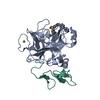
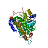
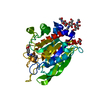
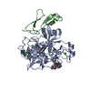
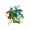
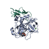
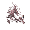
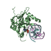
- PDB-3qi5: Crystal structure of human alkyladenine DNA glycosylase in comple... -
+
Open data
ID or keywords:
Loading...
-
Basic information
Entry
Database: PDB / ID: 3qi5
Title
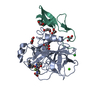
Crystal structure of human alkyladenine DNA glycosylase in complex with 3,N4-ethenocystosine containing duplex DNA
Components
DNA (5'-D(*GP*AP*CP*AP*TP*GP*(EDC)P*TP*TP*GP*CP*CP*T)-3')
DNA (5'-D(*GP*GP*CP*AP*AP*GP*CP*AP*TP*GP*TP*CP*A)-3')
DNA-3-methyladenine glycosylase
Keywords
HYDROLASE/DNA / alkyladenine DNA glycosylase fold / AAG / Excision / DNA repair / DNA binding / Nucleus / HYDROLASE-DNA complex
Function / homology
Function and homology information
alkylbase DNA N-glycosylase activity / DNA-3-methyladenine glycosylase II / depurination / DNA N-glycosylase activity / Displacement of DNA glycosylase by APEX1 / DNA alkylation repair / mitochondrial nucleoid / Recognition and association of DNA glycosylase with site containing an affected purine / Cleavage of the damaged purine / base-excision repair ...alkylbase DNA N-glycosylase activity / DNA-3-methyladenine glycosylase II / depurination / DNA N-glycosylase activity / Displacement of DNA glycosylase by APEX1 / DNA alkylation repair / mitochondrial nucleoid / Recognition and association of DNA glycosylase with site containing an affected purine / Cleavage of the damaged purine / base-excision repair / damaged DNA binding / mitochondrion / DNA binding / nucleoplasm / cytosol Similarity search - Function
A: DNA-3-methyladenine glycosylase B: DNA-3-methyladenine glycosylase C: DNA (5'-D(*GP*AP*CP*AP*TP*GP*(EDC)P*TP*TP*GP*CP*CP*T)-3') D: DNA (5'-D(*GP*GP*CP*AP*AP*GP*CP*AP*TP*GP*TP*CP*A)-3') E: DNA (5'-D(*GP*AP*CP*AP*TP*GP*(EDC)P*TP*TP*GP*CP*CP*T)-3') F: DNA (5'-D(*GP*GP*CP*AP*AP*GP*CP*AP*TP*GP*TP*CP*A)-3') hetero molecules
Mass: 18.015 Da / Num. of mol.: 232 / Source method: isolated from a natural source / Formula: H2O
-
Experimental details
-
Experiment
Experiment
Method: X-RAY DIFFRACTION / Number of used crystals: 1
-
Sample preparation
Crystal
Density Matthews: 2.14 Å3/Da / Density % sol: 42.41 %
Crystal grow
Temperature: 295 K / Method: vapor diffusion, hanging drop Details: The 3,N4-ethenocytosine (EDC) containing DNA duplex was prepared by annealing the EDC containing 13-mer crystallization oligonucleotide ('5-GAC ATG (EDC)TT GCC T-3') with its complementary ...Details: The 3,N4-ethenocytosine (EDC) containing DNA duplex was prepared by annealing the EDC containing 13-mer crystallization oligonucleotide ('5-GAC ATG (EDC)TT GCC T-3') with its complementary strand that contained G opposite EDC (5'-GGC AAG CAT GTC A-3'). The delta79AAG-EDC complexes were prepared by mixing equimolar ratios of delta79AAG and EDC:G 13-mer DNA duplex at the final protein-DNA complex concentration of 0.3 mM in the complex buffer (20 mM Hepes-NaOH pH 7.5, 100 mM NaCl, 0.1 mM EDTA, 5% v/v glycerol and 1 mM DTT). The complex was incubated on ice for 15 min and used for crystallization. The crystals were obtained upon mixing 1 uL of complex and 1 ul of the reservoir solution (100 mM sodium cacodylate pH 6.0, 200 mM manganese chloride and 20% polyethylene glycol (PEG)-3350) over 0.5 ml of the reservoir solution, followed by incubation for 2 days, VAPOR DIFFUSION, HANGING DROP, temperature 295K
-
Data collection
Diffraction
Mean temperature: 100 K
Diffraction source
Source: SYNCHROTRON / Site: ALS / Beamline: 12.3.1 / Wavelength: 1.116 Å
Resolution: 2.2→81 Å / Cor.coef. Fo:Fc: 0.92 / Cor.coef. Fo:Fc free: 0.887 / SU B: 15.824 / SU ML: 0.205 / Cross valid method: THROUGHOUT / ESU R Free: 0.274 / Stereochemistry target values: MAXIMUM LIKELIHOOD / Details: HYDROGENS HAVE BEEN ADDED IN THE RIDING POSITIONS
Rfactor
Num. reflection
% reflection
Selection details
Rfree
0.28427
1331
5.1 %
RANDOM
Rwork
0.23637
-
-
-
obs
0.23881
24947
96.22 %
-
all
-
24947
-
-
Solvent computation
Ion probe radii: 0.8 Å / Shrinkage radii: 0.8 Å / VDW probe radii: 1.4 Å / Solvent model: MASK
Displacement parameters
Biso mean: 15.654 Å2
Baniso -1
Baniso -2
Baniso -3
1-
0.62 Å2
0.29 Å2
0.21 Å2
2-
-
-1.06 Å2
0.13 Å2
3-
-
-
0.39 Å2
Refinement step
Cycle: LAST / Resolution: 2.2→81 Å
Protein
Nucleic acid
Ligand
Solvent
Total
Num. atoms
3249
1024
2
232
4507
Refine LS restraints
Refine-ID
Type
Dev ideal
Dev ideal target
Number
X-RAY DIFFRACTION
r_bond_refined_d
0.005
0.022
4468
X-RAY DIFFRACTION
r_angle_refined_deg
1.029
2.269
6254
X-RAY DIFFRACTION
r_dihedral_angle_1_deg
4.709
5
412
X-RAY DIFFRACTION
r_dihedral_angle_2_deg
29.351
21.895
153
X-RAY DIFFRACTION
r_dihedral_angle_3_deg
15.213
15
563
X-RAY DIFFRACTION
r_dihedral_angle_4_deg
13.988
15
44
X-RAY DIFFRACTION
r_chiral_restr
0.058
0.2
682
X-RAY DIFFRACTION
r_gen_planes_refined
0.004
0.021
3078
X-RAY DIFFRACTION
r_mcbond_it
0.486
1.5
2070
X-RAY DIFFRACTION
r_mcangle_it
0.888
2
3310
X-RAY DIFFRACTION
r_scbond_it
0.57
3
2398
X-RAY DIFFRACTION
r_scangle_it
0.999
4.5
2944
Refine LS restraints NCS
Refine-ID: X-RAY DIFFRACTION
Ens-ID
Dom-ID
Auth asym-ID
Number
Type
Rms dev position (Å)
Weight position
1
1
A
1152
tightpositional
0.29
0.05
1
1
B
184
mediumpositional
0.15
0.5
2
2
C
263
mediumpositional
0.33
0.5
2
2
E
245
mediumpositional
0.13
0.5
1
1
A
1152
tightthermal
0.08
0.5
1
1
B
184
mediumthermal
0.07
2
2
2
D
263
mediumthermal
0.14
2
2
2
F
245
mediumthermal
0.1
2
LS refinement shell
Resolution: 2.2→2.252 Å / Total num. of bins used: 20
Rfactor
Num. reflection
% reflection
Rfree
0.348
86
-
Rwork
0.289
1734
-
obs
-
-
89.61 %
Refinement TLS params.
Method: refined / Refine-ID: X-RAY DIFFRACTION
ID
L11 (°2)
L12 (°2)
L13 (°2)
L22 (°2)
L23 (°2)
L33 (°2)
S11 (Å °)
S12 (Å °)
S13 (Å °)
S21 (Å °)
S22 (Å °)
S23 (Å °)
S31 (Å °)
S32 (Å °)
S33 (Å °)
T11 (Å2)
T12 (Å2)
T13 (Å2)
T22 (Å2)
T23 (Å2)
T33 (Å2)
Origin x (Å)
Origin y (Å)
Origin z (Å)
1
2.1497
-1.1267
0.7034
2.4143
-1.1749
2.6896
0.0758
0.1762
0.0561
0.0858
-0.1194
-0.0937
0.0805
-0.0006
0.0436
0.1644
-0.0144
-0.0017
0.0189
0.0031
0.0503
19.5344
10.0103
-41.3271
2
2.3455
-1.2472
-1.2225
2.4382
1.0247
2.6073
-0.1419
0.1152
-0.0961
0.1284
0.0791
0.0643
-0.0568
0.0834
0.0628
0.0225
-0.0293
0.0057
0.2082
-0.0012
0.0578
-0.229
-9.7377
-67.7282
3
0.3987
-0.4108
-0.398
1.9566
0.3612
2.2433
0.0381
-0.0323
-0.1267
0.3619
-0.0794
0.0891
-0.0837
0.0183
0.0414
0.2316
-0.0348
0.0108
0.0233
-0.0008
0.0706
14.8238
5.6987
-21.1756
4
1.6315
-1.2689
-0.9148
7.5473
0.5121
3.3317
0.0261
-0.0565
-0.0623
0.3992
-0.2245
-0.1075
0.1154
-0.1552
0.1984
0.1557
-0.0239
-0.0134
0.0524
0.0305
0.059
15.078
4.0514
-21.6272
5
0.3443
0.26
-0.841
1.46
-0.6971
2.8385
-0.1782
0.155
-0.0593
-0.1711
0.0208
-0.0013
0.1245
-0.2473
0.1574
0.1656
-0.0765
-0.0055
0.2863
-0.0215
0.0662
4.2907
-4.9353
-87.7975
6
9.9932
-1.2419
-0.5354
0.8286
-1.2579
2.6352
0.1526
0.6552
-0.3351
-0.023
-0.1528
0.0544
-0.0159
0.1021
0.0002
0.084
-0.0081
0.0141
0.1335
-0.0303
0.0914
5.9779
-5.3712
-87.3452
Refinement TLS group
ID
Refine-ID
Refine TLS-ID
Auth asym-ID
Auth seq-ID
1
X-RAY DIFFRACTION
1
A
86 - 198
2
X-RAY DIFFRACTION
1
A
211 - 244
3
X-RAY DIFFRACTION
1
A
256 - 263
4
X-RAY DIFFRACTION
1
A
274 - 289
5
X-RAY DIFFRACTION
2
B
86 - 198
6
X-RAY DIFFRACTION
2
B
211 - 244
7
X-RAY DIFFRACTION
2
B
256 - 263
8
X-RAY DIFFRACTION
2
B
274 - 289
9
X-RAY DIFFRACTION
3
C
1 - 13
10
X-RAY DIFFRACTION
4
D
14 - 25
11
X-RAY DIFFRACTION
5
E
1 - 13
12
X-RAY DIFFRACTION
6
F
14 - 25
+
About Yorodumi
-
News
-
Feb 9, 2022. New format data for meta-information of EMDB entries
New format data for meta-information of EMDB entries
Version 3 of the EMDB header file is now the official format.
The previous official version 1.9 will be removed from the archive.
In the structure databanks used in Yorodumi, some data are registered as the other names, "COVID-19 virus" and "2019-nCoV". Here are the details of the virus and the list of structure data.
Jan 31, 2019. EMDB accession codes are about to change! (news from PDBe EMDB page)
EMDB accession codes are about to change! (news from PDBe EMDB page)
The allocation of 4 digits for EMDB accession codes will soon come to an end. Whilst these codes will remain in use, new EMDB accession codes will include an additional digit and will expand incrementally as the available range of codes is exhausted. The current 4-digit format prefixed with “EMD-” (i.e. EMD-XXXX) will advance to a 5-digit format (i.e. EMD-XXXXX), and so on. It is currently estimated that the 4-digit codes will be depleted around Spring 2019, at which point the 5-digit format will come into force.
The EM Navigator/Yorodumi systems omit the EMD- prefix.
Related info.:Q: What is EMD? / ID/Accession-code notation in Yorodumi/EM Navigator
Yorodumi is a browser for structure data from EMDB, PDB, SASBDB, etc.
This page is also the successor to EM Navigator detail page, and also detail information page/front-end page for Omokage search.
The word "yorodu" (or yorozu) is an old Japanese word meaning "ten thousand". "mi" (miru) is to see.
Related info.:EMDB / PDB / SASBDB / Comparison of 3 databanks / Yorodumi Search / Aug 31, 2016. New EM Navigator & Yorodumi / Yorodumi Papers / Jmol/JSmol / Function and homology information / Changes in new EM Navigator and Yorodumi
 Movie
Movie Controller

 Yorodumi
 Open data
 Basic information
 Components
 Keywords
 Function and homology information
 Homo sapiens (human)
 X-RAY DIFFRACTION /
  Authors
 Citation
 Structure visualization
 Downloads & links
 Other downloads
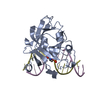

 PDBj
PDBj


 Assembly
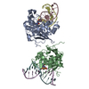


 Mass: 54.938 Da / Num. of mol.: 2 / Source method: obtained synthetically / Formula: Mn
 Mass: 18.015 Da / Num. of mol.: 232 / Source method: isolated from a natural source / Formula: H2O
 Sample preparation
 / Beamline: 12.3.1 / Wavelength: 1.116 Å
 Processing