- PDB-3uby: Crystal structure of human alklyadenine DNA glycosylase in a lowe... -
+
Open data
ID or keywords:
Loading...
-
Basic information
Entry
Database: PDB / ID: 3uby
Title
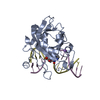
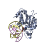
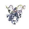
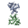
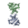
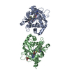
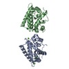
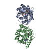
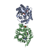
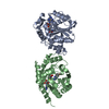
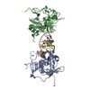
Crystal structure of human alklyadenine DNA glycosylase in a lower and higher-affinity complex with DNA
Components
DNA (5'-D(*GP*AP*CP*AP*TP*GP*(EDC)P*TP*TP*GP*CP*CP*T)-3')
DNA-3-methyladenine glycosylase
Keywords
HYDROLASE/DNA / alkyladenine DNA glycosylase fold / AAG / DNA repair / DNA binding / Nucleus / HYDROLASE-DNA complex
Function / homology
Function and homology information
alkylbase DNA N-glycosylase activity / DNA-3-methyladenine glycosylase II / depurination / DNA N-glycosylase activity / Displacement of DNA glycosylase by APEX1 / DNA alkylation repair / mitochondrial nucleoid / Recognition and association of DNA glycosylase with site containing an affected purine / Cleavage of the damaged purine / base-excision repair ...alkylbase DNA N-glycosylase activity / DNA-3-methyladenine glycosylase II / depurination / DNA N-glycosylase activity / Displacement of DNA glycosylase by APEX1 / DNA alkylation repair / mitochondrial nucleoid / Recognition and association of DNA glycosylase with site containing an affected purine / Cleavage of the damaged purine / base-excision repair / damaged DNA binding / mitochondrion / DNA binding / nucleoplasm / cytosol Similarity search - Function
A: DNA-3-methyladenine glycosylase C: DNA (5'-D(*GP*AP*CP*AP*TP*GP*(EDC)P*TP*TP*GP*CP*CP*T)-3') D: DNA (5'-D(*GP*AP*CP*AP*TP*GP*(EDC)P*TP*TP*GP*CP*CP*T)-3')
Mass: 18.015 Da / Num. of mol.: 250 / Source method: isolated from a natural source / Formula: H2O
-
Experimental details
-
Experiment
Experiment
Method: X-RAY DIFFRACTION / Number of used crystals: 1
-
Sample preparation
Crystal
Density Matthews: 1.97 Å3/Da / Density % sol: 37.46 %
Crystal grow
Temperature: 298 K / Method: vapor diffusion, hanging drop Details: An equimolar ratio of delta79AAG and 13-mer single-stranded (ss) EDC DNA were mixed to form a protein-DNA complex concentration of 0.3 mM in the complex buffer (20 mM HEPES-NaOH, pH 7.5, 100 ...Details: An equimolar ratio of delta79AAG and 13-mer single-stranded (ss) EDC DNA were mixed to form a protein-DNA complex concentration of 0.3 mM in the complex buffer (20 mM HEPES-NaOH, pH 7.5, 100 mM NaCl, 0.1 mM EDTA, 5% v/v glycerol and 1 mM DTT). The complex was incubated on ice for 15 min and used for crystallization. Crystals were obtained upon mixing 1 uL of protein-DNA complex and 1 uL of reservoir solution (100 mM BIS-TRIS, pH 5.5, 200 mM cesium chloride and 20% polyethylene glycol (PEG) 3350) over 0.5 ml of reservoir solution. Crystals appeared after incubation for 14 days at 22 degrees C, VAPOR DIFFUSION, HANGING DROP, temperature 298K
-
Data collection
Diffraction
Mean temperature: 100 K
Diffraction source
Source: SYNCHROTRON / Site: ALS / Beamline: 12.3.1 / Wavelength: 1.116 Å
Resolution: 2→65.65 Å / Cor.coef. Fo:Fc: 0.926 / Cor.coef. Fo:Fc free: 0.904 / SU B: 12.087 / SU ML: 0.158 / Cross valid method: THROUGHOUT / ESU R Free: 0.208 / Stereochemistry target values: MAXIMUM LIKELIHOOD / Details: HYDROGENS HAVE BEEN ADDED IN THE RIDING POSITIONS
Rfactor
Num. reflection
% reflection
Selection details
Rfree
0.26545
1375
5.1 %
RANDOM
Rwork
0.2194
-
-
-
obs
0.22176
25556
92.12 %
-
all
-
26998
-
-
Solvent computation
Ion probe radii: 0.8 Å / Shrinkage radii: 0.8 Å / VDW probe radii: 1.4 Å / Solvent model: MASK
Displacement parameters
Biso mean: 36.01 Å2
Baniso -1
Baniso -2
Baniso -3
1-
0.41 Å2
0 Å2
0 Å2
2-
-
0.41 Å2
0 Å2
3-
-
-
-0.82 Å2
Refinement step
Cycle: LAST / Resolution: 2→65.65 Å
Protein
Nucleic acid
Ligand
Solvent
Total
Num. atoms
2967
354
0
250
3571
Refine LS restraints
Refine-ID
Type
Dev ideal
Dev ideal target
Number
X-RAY DIFFRACTION
r_bond_refined_d
0.007
0.022
3429
X-RAY DIFFRACTION
r_angle_refined_deg
1.104
2.112
4709
X-RAY DIFFRACTION
r_dihedral_angle_1_deg
5.055
5
376
X-RAY DIFFRACTION
r_dihedral_angle_2_deg
31.096
22.029
138
X-RAY DIFFRACTION
r_dihedral_angle_3_deg
14.599
15
504
X-RAY DIFFRACTION
r_dihedral_angle_4_deg
14.494
15
38
X-RAY DIFFRACTION
r_chiral_restr
0.068
0.2
513
X-RAY DIFFRACTION
r_gen_planes_refined
0.005
0.021
2502
X-RAY DIFFRACTION
r_mcbond_it
2.61
1.5
1900
X-RAY DIFFRACTION
r_mcangle_it
3.499
2
3031
X-RAY DIFFRACTION
r_scbond_it
5.26
3
1529
X-RAY DIFFRACTION
r_scangle_it
6.684
4.5
1678
LS refinement shell
Resolution: 2→2.053 Å / Total num. of bins used: 20
Rfactor
Num. reflection
% reflection
Rfree
0.265
99
-
Rwork
0.219
1833
-
obs
-
-
89.49 %
Refinement TLS params.
Method: refined / Refine-ID: X-RAY DIFFRACTION
ID
L11 (°2)
L12 (°2)
L13 (°2)
L22 (°2)
L23 (°2)
L33 (°2)
S11 (Å °)
S12 (Å °)
S13 (Å °)
S21 (Å °)
S22 (Å °)
S23 (Å °)
S31 (Å °)
S32 (Å °)
S33 (Å °)
T11 (Å2)
T12 (Å2)
T13 (Å2)
T22 (Å2)
T23 (Å2)
T33 (Å2)
Origin x (Å)
Origin y (Å)
Origin z (Å)
1
1.8175
-0.756
-0.8565
1.83
0.6659
2.5151
0.0444
0.0808
-0.0785
0.1421
-0.1046
0.0499
-0.0796
0.0603
0.0602
0.2203
0.0217
-0.0121
0.0209
-0.0225
0.1267
3.6984
-5.0095
21.4544
2
1.7615
-0.874
0.8017
3.1614
-0.6637
3.6281
-0.0657
0.184
0.0425
-0.1014
0.1151
-0.0269
0.0369
-0.1413
-0.0493
0.1001
0.0275
0.0268
0.1592
-0.0474
0.1279
-15.1881
-26.4475
-1.8766
3
0.5437
0.9939
1.202
1.8201
2.2202
2.838
0.0469
-0.0752
0.0119
0.1083
-0.1109
0.0343
0.1071
0.1185
0.0641
0.4785
-0.0197
-0.025
0.4773
0.0084
0.218
7.7356
1.8047
40.2028
4
1.8467
0.513
-2.3749
10.8793
-2.1692
12.9283
0.0918
-0.5275
0.355
0.1186
0.4908
-0.4344
-0.3425
0.3405
-0.5826
0.2237
-0.0591
-0.0267
0.2337
-0.1381
0.1711
9.5881
9.3631
38.6698
Refinement TLS group
ID
Refine-ID
Refine TLS-ID
Auth asym-ID
Auth seq-ID
1
X-RAY DIFFRACTION
1
A
80 - 200
2
X-RAY DIFFRACTION
1
A
209 - 264
3
X-RAY DIFFRACTION
1
A
268 - 290
4
X-RAY DIFFRACTION
2
B
80 - 130
5
X-RAY DIFFRACTION
2
B
144 - 160
6
X-RAY DIFFRACTION
2
B
166 - 198
7
X-RAY DIFFRACTION
2
B
208 - 262
8
X-RAY DIFFRACTION
2
B
274 - 289
9
X-RAY DIFFRACTION
3
C
1 - 11
10
X-RAY DIFFRACTION
4
D
1 - 8
+
About Yorodumi
-
News
-
Feb 9, 2022. New format data for meta-information of EMDB entries
New format data for meta-information of EMDB entries
Version 3 of the EMDB header file is now the official format.
The previous official version 1.9 will be removed from the archive.
In the structure databanks used in Yorodumi, some data are registered as the other names, "COVID-19 virus" and "2019-nCoV". Here are the details of the virus and the list of structure data.
Jan 31, 2019. EMDB accession codes are about to change! (news from PDBe EMDB page)
EMDB accession codes are about to change! (news from PDBe EMDB page)
The allocation of 4 digits for EMDB accession codes will soon come to an end. Whilst these codes will remain in use, new EMDB accession codes will include an additional digit and will expand incrementally as the available range of codes is exhausted. The current 4-digit format prefixed with “EMD-” (i.e. EMD-XXXX) will advance to a 5-digit format (i.e. EMD-XXXXX), and so on. It is currently estimated that the 4-digit codes will be depleted around Spring 2019, at which point the 5-digit format will come into force.
The EM Navigator/Yorodumi systems omit the EMD- prefix.
Related info.:Q: What is EMD? / ID/Accession-code notation in Yorodumi/EM Navigator
Yorodumi is a browser for structure data from EMDB, PDB, SASBDB, etc.
This page is also the successor to EM Navigator detail page, and also detail information page/front-end page for Omokage search.
The word "yorodu" (or yorozu) is an old Japanese word meaning "ten thousand". "mi" (miru) is to see.
Related info.:EMDB / PDB / SASBDB / Comparison of 3 databanks / Yorodumi Search / Aug 31, 2016. New EM Navigator & Yorodumi / Yorodumi Papers / Jmol/JSmol / Function and homology information / Changes in new EM Navigator and Yorodumi
 Movie
Movie Controller

 Yorodumi
 Open data
 Basic information
 Components
 Keywords
 Function and homology information
 Homo sapiens (human)
 X-RAY DIFFRACTION /
  Authors
 Citation
 Structure visualization
 Downloads & links
 Other downloads
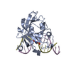


 PDBj
PDBj


 Assembly

 Mass: 18.015 Da / Num. of mol.: 250 / Source method: isolated from a natural source / Formula: H2O
 Sample preparation
 / Beamline: 12.3.1 / Wavelength: 1.116 Å
 Processing