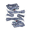
 ムービー
ムービー コントローラー


+ データを開く

- 基本情報
基本情報
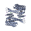
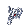
| 登録情報 | データベース: PDB / ID: 3ual | ||||||
|---|---|---|---|---|---|---|---|
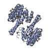
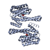
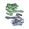
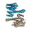
| タイトル | Crystal Structure of 14-3-3 epsilon with Mlf1 peptide | ||||||
 要素 要素 |
| ||||||
 キーワード キーワード | SIGNALING PROTEIN/PROTEIN BINDING / Adapter protein / ALL HELICAL / PHOSPHOPETIDE / SIGNALING PROTEIN-PROTEIN BINDING complex | ||||||
| 機能・相同性 |  機能・相同性情報 機能・相同性情報regulation of heart rate by hormone / positive regulation of hippo signaling / membrane repolarization during cardiac muscle cell action potential / regulation of cell cycle G1/S phase transition / protein localization to endoplasmic reticulum / negative regulation of toll-like receptor signaling pathway / regulation of membrane repolarization / NADE modulates death signalling / RAB GEFs exchange GTP for GDP on RABs / myeloid progenitor cell differentiation ...regulation of heart rate by hormone / positive regulation of hippo signaling / membrane repolarization during cardiac muscle cell action potential / regulation of cell cycle G1/S phase transition / protein localization to endoplasmic reticulum / negative regulation of toll-like receptor signaling pathway / regulation of membrane repolarization / NADE modulates death signalling / RAB GEFs exchange GTP for GDP on RABs / myeloid progenitor cell differentiation / Signaling by Hippo / regulation of potassium ion transmembrane transport / protein phosphatase inhibitor activity / cytoplasmic pattern recognition receptor signaling pathway / Deregulated CDK5 triggers multiple neurodegenerative pathways in Alzheimer's disease models / negative regulation of calcium ion export across plasma membrane / intracellular potassium ion homeostasis / regulation of heart rate by cardiac conduction / Regulation of HSF1-mediated heat shock response / Activation of BAD and translocation to mitochondria / phosphoserine residue binding / protein localization to nucleus / regulation of cytosolic calcium ion concentration / HSF1 activation / potassium channel regulator activity / protein targeting / Chk1/Chk2(Cds1) mediated inactivation of Cyclin B:Cdk1 complex / SARS-CoV-2 targets host intracellular signalling and regulatory pathways / calcium channel inhibitor activity / RHO GTPases activate PKNs / SARS-CoV-1 targets host intracellular signalling and regulatory pathways / signaling adaptor activity / Loss of Nlp from mitotic centrosomes / Loss of proteins required for interphase microtubule organization from the centrosome / Transcriptional and post-translational regulation of MITF-M expression and activity / Recruitment of mitotic centrosome proteins and complexes / substantia nigra development / Recruitment of NuMA to mitotic centrosomes / Anchoring of the basal body to the plasma membrane / protein sequestering activity / regulation of mitotic cell cycle / regulation of signal transduction by p53 class mediator / AURKA Activation by TPX2 / positive regulation of protein export from nucleus / TP53 Regulates Metabolic Genes / Translocation of SLC2A4 (GLUT4) to the plasma membrane / calcium channel regulator activity / hippocampus development / phosphoprotein binding / cerebral cortex development / mitochondrial membrane / histone deacetylase binding / neuron migration / MHC class II protein complex binding / Regulation of PLK1 Activity at G2/M Transition / intracellular protein localization / melanosome / MAPK cascade / cellular response to heat / scaffold protein binding / protein phosphatase binding / transmembrane transporter binding / intracellular signal transduction / cilium / ciliary basal body / cadherin binding / protein heterodimerization activity / protein domain specific binding / focal adhesion / DNA-templated transcription / ubiquitin protein ligase binding / regulation of DNA-templated transcription / perinuclear region of cytoplasm / enzyme binding / endoplasmic reticulum / signal transduction / DNA binding / RNA binding / extracellular exosome / identical protein binding / nucleus / membrane / cytosol / cytoplasm 類似検索 - 分子機能 | ||||||
| 生物種 |  Homo sapiens (ヒト) Homo sapiens (ヒト) | ||||||
| 手法 |  X線回折 / シンクロトロン / 分子置換 / 解像度: 1.8 Å X線回折 / シンクロトロン / 分子置換 / 解像度: 1.8 Å | ||||||
 データ登録者 データ登録者 | Weyand, M. / Ottmann, C. | ||||||
 引用 引用 | ジャーナル: Febs J. / 年: 2012 タイトル: Structural insights of the MLF1/14-3-3 interaction. 著者: Molzan, M. / Weyand, M. / Rose, R. / Ottmann, C. | ||||||
| 履歴 |
|
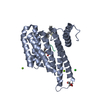
- 構造の表示
構造の表示
| 構造ビューア | 分子: MolmilJmol/JSmol |
|---|
- ダウンロードとリンク
-ダウンロード
| PDBx/mmCIF形式 | 3ual.cif.gz | 117.5 KB | 表示 | PDBx/mmCIF形式 |
|---|---|---|---|---|
| PDB形式 | pdb3ual.ent.gz | 92.1 KB | 表示 | PDB形式 |
| PDBx/mmJSON形式 | 3ual.json.gz | ツリー表示 | PDBx/mmJSON形式 | |
| その他 |  その他のダウンロード その他のダウンロード |
-検証レポート
| 文書・要旨 | 3ual_validation.pdf.gz | 448.6 KB | 表示 | wwPDB検証レポート |
|---|---|---|---|---|
| 文書・詳細版 | 3ual_full_validation.pdf.gz | 450.3 KB | 表示 | |
| XML形式データ | 3ual_validation.xml.gz | 13.1 KB | 表示 | |
| CIF形式データ | 3ual_validation.cif.gz | 18.4 KB | 表示 | |
| アーカイブディレクトリ | https://data.pdbj.org/pub/pdb/validation_reports/ua/3ualftp://data.pdbj.org/pub/pdb/validation_reports/ua/3ual | HTTPS FTP |
-関連構造データ


-リンク
 PDBj
PDBj


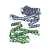
- 集合体
集合体
| 登録構造単位 | 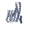
| ||||||||
|---|---|---|---|---|---|---|---|---|---|
| 1 | 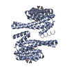
| ||||||||
| 2 |
| ||||||||
| 単位格子 |
| ||||||||
| Components on special symmetry positions |
|
-要素
| #1: タンパク質 | 分子量: 26617.340 Da / 分子数: 1 / 断片: UNP residues 1-232 / 由来タイプ: 組換発現 / 由来: (組換発現) Homo sapiens (ヒト) / 遺伝子: YWHAE / 発現宿主:  | ||||
|---|---|---|---|---|---|
| #2: タンパク質・ペプチド | 分子量: 1749.922 Da / 分子数: 1 / 断片: UNP residues 29-42 / 由来タイプ: 合成 / 詳細: This sequence occurs naturally in humans. / 由来: (合成) Homo sapiens (ヒト) / 参照: UniProt: P58340 | ||||
| #3: 化合物 | 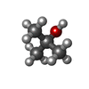  分子量: 74.122 Da / 分子数: 3 / 由来タイプ: 合成 / 式: C4H10O 分子量: 74.122 Da / 分子数: 3 / 由来タイプ: 合成 / 式: C4H10O#4: 水 | ChemComp-HOH / |  分子量: 18.015 Da / 分子数: 159 / 由来タイプ: 天然 / 式: H2O 分子量: 18.015 Da / 分子数: 159 / 由来タイプ: 天然 / 式: H2OHas protein modification | Y | |
-実験情報
-実験
| 実験 | 手法: X線回折 / 使用した結晶の数: 1 |
|---|
- 試料調製
試料調製
| 結晶 | マシュー密度: 2.32 Å3/Da / 溶媒含有率: 46.95 % |
|---|---|
| 結晶化 | 温度: 277 K / 手法: 蒸気拡散法, ハンギングドロップ法 / pH: 5.6 詳細: 0.1 M Na-Citrate, 35% tert-butanol, pH 5.6, VAPOR DIFFUSION, HANGING DROP, temperature 277K |
-データ収集
| 回折 | 平均測定温度: 100 K |
|---|---|
| 放射光源 | 由来: シンクロトロン / サイト: SLS  / ビームライン: X10SA / 波長: 0.97906 Å / ビームライン: X10SA / 波長: 0.97906 Å |
| 検出器 | タイプ: MARMOSAIC 225 mm CCD / 検出器: CCD / 日付: 2008年1月25日 |
| 放射 | モノクロメーター: LN2 cooled fixed-exit Si(111) monochromator プロトコル: SINGLE WAVELENGTH / 単色(M)・ラウエ(L): M / 散乱光タイプ: x-ray |
| 放射波長 | 波長: 0.97906 Å / 相対比: 1 |
| 反射 | 解像度: 1.8→100 Å / Num. all: 24815 / Num. obs: 24289 / % possible obs: 97.9 % / Observed criterion σ(I): -3 / 冗長度: 6.1 % / Biso Wilson estimate: 32.9 Å2 / Rmerge(I) obs: 0.068 / Rsym value: 0.061 / Net I/σ(I): 16.8 |
| 反射 シェル | 解像度: 1.8→1.9 Å / 冗長度: 6.2 % / Rmerge(I) obs: 0.456 / Mean I/σ(I) obs: 4.1 / Num. unique all: 3568 / Rsym value: 0.45 / % possible all: 97.6 |
- 解析
解析
| ソフトウェア |
| ||||||||||||||||||||||||||||||||||||||||||||||||||||||||||||||||||||||||||||||||||||||||||||||||||||||||||||||||||||||||||||||||||||||||||||||||||||||||||||||||||||||||||
|---|---|---|---|---|---|---|---|---|---|---|---|---|---|---|---|---|---|---|---|---|---|---|---|---|---|---|---|---|---|---|---|---|---|---|---|---|---|---|---|---|---|---|---|---|---|---|---|---|---|---|---|---|---|---|---|---|---|---|---|---|---|---|---|---|---|---|---|---|---|---|---|---|---|---|---|---|---|---|---|---|---|---|---|---|---|---|---|---|---|---|---|---|---|---|---|---|---|---|---|---|---|---|---|---|---|---|---|---|---|---|---|---|---|---|---|---|---|---|---|---|---|---|---|---|---|---|---|---|---|---|---|---|---|---|---|---|---|---|---|---|---|---|---|---|---|---|---|---|---|---|---|---|---|---|---|---|---|---|---|---|---|---|---|---|---|---|---|---|---|---|---|
| 精密化 | 構造決定の手法: 分子置換 開始モデル: PDB ENTRY 2BR9 解像度: 1.8→35.27 Å / Cor.coef. Fo:Fc: 0.964 / Cor.coef. Fo:Fc free: 0.937 / SU B: 4.996 / SU ML: 0.077 / 交差検証法: THROUGHOUT / σ(F): 0 / ESU R Free: 0.125 / 立体化学のターゲット値: MAXIMUM LIKELIHOOD / 詳細: HYDROGENS HAVE BEEN ADDED IN THE RIDING POSITIONS
| ||||||||||||||||||||||||||||||||||||||||||||||||||||||||||||||||||||||||||||||||||||||||||||||||||||||||||||||||||||||||||||||||||||||||||||||||||||||||||||||||||||||||||
| 溶媒の処理 | イオンプローブ半径: 0.8 Å / 減衰半径: 0.8 Å / VDWプローブ半径: 1.2 Å / 溶媒モデル: MASK | ||||||||||||||||||||||||||||||||||||||||||||||||||||||||||||||||||||||||||||||||||||||||||||||||||||||||||||||||||||||||||||||||||||||||||||||||||||||||||||||||||||||||||
| 原子変位パラメータ | Biso mean: 32.07 Å2
| ||||||||||||||||||||||||||||||||||||||||||||||||||||||||||||||||||||||||||||||||||||||||||||||||||||||||||||||||||||||||||||||||||||||||||||||||||||||||||||||||||||||||||
| 精密化ステップ | サイクル: LAST / 解像度: 1.8→35.27 Å
| ||||||||||||||||||||||||||||||||||||||||||||||||||||||||||||||||||||||||||||||||||||||||||||||||||||||||||||||||||||||||||||||||||||||||||||||||||||||||||||||||||||||||||
| 拘束条件 |
| ||||||||||||||||||||||||||||||||||||||||||||||||||||||||||||||||||||||||||||||||||||||||||||||||||||||||||||||||||||||||||||||||||||||||||||||||||||||||||||||||||||||||||
| LS精密化 シェル | 解像度: 1.8→1.847 Å / Total num. of bins used: 20
| ||||||||||||||||||||||||||||||||||||||||||||||||||||||||||||||||||||||||||||||||||||||||||||||||||||||||||||||||||||||||||||||||||||||||||||||||||||||||||||||||||||||||||
| 精密化 TLS | 手法: refined / Origin x: -18.8512 Å / Origin y: 5.6095 Å / Origin z: 20.9416 Å
|
