 Movie
Movie Controller

[English] 日本語
 Yorodumi
- PDB-3uai: Structure of the Shq1-Cbf5-Nop10-Gar1 complex from Saccharomyces ... -
+ Open data

- Basic information
| Entry | Database: PDB / ID: 3uai | ||||||
|---|---|---|---|---|---|---|---|
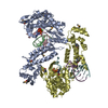
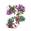
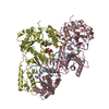
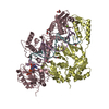
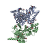
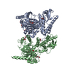
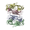
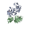
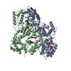
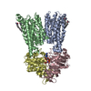
| Title | Structure of the Shq1-Cbf5-Nop10-Gar1 complex from Saccharomyces cerevisiae | ||||||
 Components Components |
| ||||||
 Keywords Keywords | ISOMERASE/CHAPERONE / H/ACA RNP assembly intermediate / H/ACA RNA / Nuclear / ISOMERASE-CHAPERONE complex | ||||||
| Function / homology |  Function and homology information Function and homology informationsnRNA pseudouridine synthase activity / Telomere Extension By Telomerase / box H/ACA snoRNP assembly / snoRNA guided rRNA pseudouridine synthesis / Isomerases; Intramolecular transferases; Transferring other groups / rRNA pseudouridine synthesis / box H/ACA snoRNP complex / box H/ACA sno(s)RNA 3'-end processing / snRNA pseudouridine synthesis / mRNA pseudouridine synthesis ...snRNA pseudouridine synthase activity / Telomere Extension By Telomerase / box H/ACA snoRNP assembly / snoRNA guided rRNA pseudouridine synthesis / Isomerases; Intramolecular transferases; Transferring other groups / rRNA pseudouridine synthesis / box H/ACA snoRNP complex / box H/ACA sno(s)RNA 3'-end processing / snRNA pseudouridine synthesis / mRNA pseudouridine synthesis / box H/ACA snoRNA binding / pseudouridine synthase activity / rRNA modification / sno(s)RNA-containing ribonucleoprotein complex / telomerase RNA binding / snoRNA binding / chromosome, centromeric region / 90S preribosome / rRNA processing / : / microtubule / cell division / mRNA binding / nucleolus / DNA binding / RNA binding / nucleoplasm / nucleus / cytoplasm / cytosol Similarity search - Function | ||||||
| Biological species |  | ||||||
| Method |  X-RAY DIFFRACTION / SYNCHROTRON / MOLECULAR REPLACEMENT / Resolution: 3.06 Å X-RAY DIFFRACTION / SYNCHROTRON / MOLECULAR REPLACEMENT / Resolution: 3.06 Å | ||||||
 Authors Authors | Ye, K. | ||||||
 Citation Citation | Journal: Embo J. / Year: 2011 Title: Structure of the Shq1-Cbf5-Nop10-Gar1 complex and implications for H/ACA RNP biogenesis and dyskeratosis congenita Authors: Li, S. / Duan, J. / Li, D. / Ma, S. / Ye, K. | ||||||
| History |
|
- Structure visualization
| Structure viewer | Molecule: MolmilJmol/JSmol |
|---|
- Downloads & links
-Download
| PDBx/mmCIF format | 3uai.cif.gz | 346.6 KB | Display | PDBx/mmCIF format |
|---|---|---|---|---|
| PDB format | pdb3uai.ent.gz | 285.8 KB | Display | PDB format |
| PDBx/mmJSON format | 3uai.json.gz | Tree view | PDBx/mmJSON format | |
| Others |  Other downloads Other downloads |
-Validation report
| Arichive directory | https://data.pdbj.org/pub/pdb/validation_reports/ua/3uaiftp://data.pdbj.org/pub/pdb/validation_reports/ua/3uai | HTTPS FTP |
|---|
-Related structure data
| Related structure data | 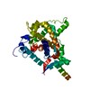 3uahSC 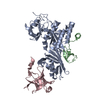 3u28S S: Starting model for refinement C: citing same article ( |
|---|---|
| Similar structure data |
-Links
 PDBj
PDBj


- Assembly
| Deposited unit | 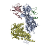
| ||||||||
|---|---|---|---|---|---|---|---|---|---|
| 1 |
| ||||||||
| Unit cell |
|
-Components
| #1: Protein | Mass: 44981.797 Da / Num. of mol.: 1 / Fragment: Core domain, UNP residues 3-394 Source method: isolated from a genetically manipulated source Source: (gene. exp.) Strain: S288c / Gene: CBF5 / Plasmid: pETDuet-1 / Production host:  References: UniProt: P33322, Isomerases; Intramolecular transferases; Transferring other groups |
|---|---|
| #2: Protein | Mass: 6649.745 Da / Num. of mol.: 1 Source method: isolated from a genetically manipulated source Source: (gene. exp.) Strain: S288c / Gene: NOP10 / Plasmid: pETDuet-1 / Production host: |
| #3: Protein | Mass: 12618.431 Da / Num. of mol.: 1 / Fragment: Core domain, UNP residues 32-124 Source method: isolated from a genetically manipulated source Source: (gene. exp.) Strain: S288c / Gene: GAR1 / Plasmid: pET28a / Production host: |
| #4: Protein | Mass: 42928.047 Da / Num. of mol.: 1 / Fragment: Shq1 specific domain, UNP residues 147-504 Source method: isolated from a genetically manipulated source Source: (gene. exp.) Strain: S288c / Gene: SHQ1 / Plasmid: pETDuet-1 / Production host: |
-Experimental details
-Experiment
| Experiment | Method: X-RAY DIFFRACTION / Number of used crystals: 1 |
|---|
- Sample preparation
| Crystal | Density Matthews: 3.24 Å3/Da / Density % sol: 62.03 % |
|---|---|
| Crystal grow | Temperature: 297 K / Method: vapor diffusion, hanging drop / pH: 6.5 Details: 0.1M Bis-Tris, 20%(w/v) polyethylene glycol monomethyl ether 5000, pH 6.5, VAPOR DIFFUSION, HANGING DROP, temperature 297K |
-Data collection
| Diffraction | Mean temperature: 100 K |
|---|---|
| Diffraction source | Source: SYNCHROTRON / Site: SSRF  / Beamline: BL17U / Wavelength: 0.9789 Å / Beamline: BL17U / Wavelength: 0.9789 Å |
| Detector | Detector: CCD / Date: Jun 6, 2011 |
| Radiation | Protocol: SINGLE WAVELENGTH / Monochromatic (M) / Laue (L): M / Scattering type: x-ray |
| Radiation wavelength | Wavelength: 0.9789 Å / Relative weight: 1 |
| Reflection | Resolution: 3.06→50 Å / Num. obs: 27487 / % possible obs: 99.9 % / Observed criterion σ(F): 0 / Observed criterion σ(I): 0 / Redundancy: 14.1 % / Rmerge(I) obs: 0.067 / Net I/σ(I): 58.4 |
| Reflection shell | Resolution: 3.06→3.11 Å / Redundancy: 14.7 % / Rmerge(I) obs: 0.684 / Mean I/σ(I) obs: 5.6 / % possible all: 100 |
- Processing
| Software |
| |||||||||||||||||||||||||||||||||||||||||||||||||||||||||||||||||||||||||||||||||||||||||||||||||||||||||||||||||||||||||||||||||||||||||||||||||||||||||||||||||||||||||||||||||||||||||||||||||||||||||||||||||||||||||||||||||
|---|---|---|---|---|---|---|---|---|---|---|---|---|---|---|---|---|---|---|---|---|---|---|---|---|---|---|---|---|---|---|---|---|---|---|---|---|---|---|---|---|---|---|---|---|---|---|---|---|---|---|---|---|---|---|---|---|---|---|---|---|---|---|---|---|---|---|---|---|---|---|---|---|---|---|---|---|---|---|---|---|---|---|---|---|---|---|---|---|---|---|---|---|---|---|---|---|---|---|---|---|---|---|---|---|---|---|---|---|---|---|---|---|---|---|---|---|---|---|---|---|---|---|---|---|---|---|---|---|---|---|---|---|---|---|---|---|---|---|---|---|---|---|---|---|---|---|---|---|---|---|---|---|---|---|---|---|---|---|---|---|---|---|---|---|---|---|---|---|---|---|---|---|---|---|---|---|---|---|---|---|---|---|---|---|---|---|---|---|---|---|---|---|---|---|---|---|---|---|---|---|---|---|---|---|---|---|---|---|---|---|---|---|---|---|---|---|---|---|---|---|---|---|---|---|---|---|
| Refinement | Method to determine structure: MOLECULAR REPLACEMENT Starting model: 3UAH, 3U28 Resolution: 3.06→20 Å / Cor.coef. Fo:Fc: 0.942 / Cor.coef. Fo:Fc free: 0.907 / SU B: 37.907 / SU ML: 0.311 / Cross valid method: THROUGHOUT / σ(F): 0 / ESU R Free: 0.434 / Stereochemistry target values: MAXIMUM LIKELIHOOD / Details: HYDROGENS HAVE BEEN ADDED IN THE RIDING POSITIONS
| |||||||||||||||||||||||||||||||||||||||||||||||||||||||||||||||||||||||||||||||||||||||||||||||||||||||||||||||||||||||||||||||||||||||||||||||||||||||||||||||||||||||||||||||||||||||||||||||||||||||||||||||||||||||||||||||||
| Solvent computation | Ion probe radii: 0.8 Å / Shrinkage radii: 0.8 Å / VDW probe radii: 1.4 Å / Solvent model: MASK | |||||||||||||||||||||||||||||||||||||||||||||||||||||||||||||||||||||||||||||||||||||||||||||||||||||||||||||||||||||||||||||||||||||||||||||||||||||||||||||||||||||||||||||||||||||||||||||||||||||||||||||||||||||||||||||||||
| Displacement parameters | Biso mean: 100.637 Å2
| |||||||||||||||||||||||||||||||||||||||||||||||||||||||||||||||||||||||||||||||||||||||||||||||||||||||||||||||||||||||||||||||||||||||||||||||||||||||||||||||||||||||||||||||||||||||||||||||||||||||||||||||||||||||||||||||||
| Refinement step | Cycle: LAST / Resolution: 3.06→20 Å
| |||||||||||||||||||||||||||||||||||||||||||||||||||||||||||||||||||||||||||||||||||||||||||||||||||||||||||||||||||||||||||||||||||||||||||||||||||||||||||||||||||||||||||||||||||||||||||||||||||||||||||||||||||||||||||||||||
| Refine LS restraints |
| |||||||||||||||||||||||||||||||||||||||||||||||||||||||||||||||||||||||||||||||||||||||||||||||||||||||||||||||||||||||||||||||||||||||||||||||||||||||||||||||||||||||||||||||||||||||||||||||||||||||||||||||||||||||||||||||||
| LS refinement shell | Resolution: 3.062→3.14 Å / Total num. of bins used: 20
| |||||||||||||||||||||||||||||||||||||||||||||||||||||||||||||||||||||||||||||||||||||||||||||||||||||||||||||||||||||||||||||||||||||||||||||||||||||||||||||||||||||||||||||||||||||||||||||||||||||||||||||||||||||||||||||||||
| Refinement TLS params. | Method: refined / Refine-ID: X-RAY DIFFRACTION
| |||||||||||||||||||||||||||||||||||||||||||||||||||||||||||||||||||||||||||||||||||||||||||||||||||||||||||||||||||||||||||||||||||||||||||||||||||||||||||||||||||||||||||||||||||||||||||||||||||||||||||||||||||||||||||||||||
| Refinement TLS group |
|
