 Movie
Movie Controller


+ Open data

- Basic information
| Entry | Database: PDB / ID: 3vxt | ||||||
|---|---|---|---|---|---|---|---|
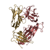
| Title | T36-5 TCR specific for HLA-A24-Nef134-10 | ||||||
 Components Components |
| ||||||
 Keywords Keywords | IMMUNE SYSTEM / HIV-1 / NEF / T CELL RECEPTOR / IMMUNOGLOBURIN DOMAIN / TCR / IMMUNE RESPONSE | ||||||
| Function / homology | Immunoglobulins / Immunoglobulin-like / Sandwich / Mainly Beta Function and homology information Function and homology information | ||||||
| Biological species |  Homo sapiens (human) Homo sapiens (human) | ||||||
| Method |  X-RAY DIFFRACTION / SYNCHROTRON / MOLECULAR REPLACEMENT / Resolution: 2.5 Å X-RAY DIFFRACTION / SYNCHROTRON / MOLECULAR REPLACEMENT / Resolution: 2.5 Å | ||||||
 Authors Authors | Shimizu, A. / Fukai, S. / Yamagata, A. / Iwamoto, A. | ||||||
 Citation Citation | Journal: SCI REP / Year: 2013 Title: Structure of TCR and antigen complexes at an immunodominant CTL epitope in HIV-1 infection Authors: Shimizu, A. / Kawana-Tachikawa, A. / Yamagata, A. / Han, C. / Zhu, D. / Sato, Y. / Nakamura, H. / Koibuchi, T. / Carlson, J. / Martin, E. / Brumme, C.J. / Shi, Y. / Gao, G.F. / Brumme, Z.L. ...Authors: Shimizu, A. / Kawana-Tachikawa, A. / Yamagata, A. / Han, C. / Zhu, D. / Sato, Y. / Nakamura, H. / Koibuchi, T. / Carlson, J. / Martin, E. / Brumme, C.J. / Shi, Y. / Gao, G.F. / Brumme, Z.L. / Fukai, S. / Iwamoto, A. | ||||||
| History |
|
- Structure visualization
| Structure viewer | Molecule: MolmilJmol/JSmol |
|---|
- Downloads & links
-Download
| PDBx/mmCIF format | 3vxt.cif.gz | 184.3 KB | Display | PDBx/mmCIF format |
|---|---|---|---|---|
| PDB format | pdb3vxt.ent.gz | 146.5 KB | Display | PDB format |
| PDBx/mmJSON format | 3vxt.json.gz | Tree view | PDBx/mmJSON format | |
| Others |  Other downloads Other downloads |
-Validation report
| Arichive directory | https://data.pdbj.org/pub/pdb/validation_reports/vx/3vxtftp://data.pdbj.org/pub/pdb/validation_reports/vx/3vxt | HTTPS FTP |
|---|
-Related structure data
| Related structure data |  3vxmC  3vxnC  3vxoC  3vxpC  3vxqC  3vxrC  3vxsC  3vxuC  3w0wC  3kxfS C: citing same article ( S: Starting model for refinement |
|---|---|
| Similar structure data |


-Links
 PDBj
PDBj
- Assembly
| Deposited unit | 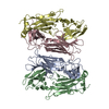
| ||||||||
|---|---|---|---|---|---|---|---|---|---|
| 1 | 
| ||||||||
| 2 | 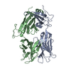
| ||||||||
| Unit cell |
|
-Components
| #1: Protein | Mass: 22764.934 Da / Num. of mol.: 2 Source method: isolated from a genetically manipulated source Source: (gene. exp.) Homo sapiens (human) / Plasmid: PET21A(+) / Production host:  #2: Protein | Mass: 27577.764 Da / Num. of mol.: 2 Source method: isolated from a genetically manipulated source Source: (gene. exp.) Homo sapiens (human) / Plasmid: PET21A(+) / Production host: #3: Water | ChemComp-HOH / |  Mass: 18.015 Da / Num. of mol.: 108 / Source method: isolated from a natural source / Formula: H2O Mass: 18.015 Da / Num. of mol.: 108 / Source method: isolated from a natural source / Formula: H2OHas protein modification | Y | |
|---|
-Experimental details
-Experiment
| Experiment | Method: X-RAY DIFFRACTION / Number of used crystals: 1 |
|---|
- Sample preparation
| Crystal | Density Matthews: 2.63 Å3/Da / Density % sol: 53.18 % / Mosaicity: 0.596 ° |
|---|---|
| Crystal grow | Temperature: 293 K / Method: vapor diffusion, sitting drop / pH: 6 Details: 0.2M potassium sulfate, 0.1M MES, 15% PEG8000, pH 6.0, VAPOR DIFFUSION, SITTING DROP, temperature 293K |
-Data collection
| Diffraction | Mean temperature: 100 K | |||||||||||||||||||||||||||||||||||||||||||||||||||||||||||||||||||||||||||||
|---|---|---|---|---|---|---|---|---|---|---|---|---|---|---|---|---|---|---|---|---|---|---|---|---|---|---|---|---|---|---|---|---|---|---|---|---|---|---|---|---|---|---|---|---|---|---|---|---|---|---|---|---|---|---|---|---|---|---|---|---|---|---|---|---|---|---|---|---|---|---|---|---|---|---|---|---|---|---|
| Diffraction source | Source: SYNCHROTRON / Site: SPring-8  / Beamline: BL41XU / Wavelength: 1 Å / Beamline: BL41XU / Wavelength: 1 Å | |||||||||||||||||||||||||||||||||||||||||||||||||||||||||||||||||||||||||||||
| Detector | Type: RAYONIX MX225HE / Detector: CCD / Date: Oct 29, 2010 | |||||||||||||||||||||||||||||||||||||||||||||||||||||||||||||||||||||||||||||
| Radiation | Protocol: SINGLE WAVELENGTH / Monochromatic (M) / Laue (L): M / Scattering type: x-ray | |||||||||||||||||||||||||||||||||||||||||||||||||||||||||||||||||||||||||||||
| Radiation wavelength | Wavelength: 1 Å / Relative weight: 1 | |||||||||||||||||||||||||||||||||||||||||||||||||||||||||||||||||||||||||||||
| Reflection | Resolution: 2.5→50 Å / Num. obs: 32447 / % possible obs: 91.9 % / Observed criterion σ(I): 0 / Redundancy: 3 % / Rsym value: 0.038 / Χ2: 1.675 / Net I/σ(I): 23.5 | |||||||||||||||||||||||||||||||||||||||||||||||||||||||||||||||||||||||||||||
| Reflection shell |
|
- Processing
| Software |
| ||||||||||||||||||||||||||||||||||||
|---|---|---|---|---|---|---|---|---|---|---|---|---|---|---|---|---|---|---|---|---|---|---|---|---|---|---|---|---|---|---|---|---|---|---|---|---|---|
| Refinement | Method to determine structure: MOLECULAR REPLACEMENT Starting model: 3KXF Resolution: 2.5→50 Å / Occupancy max: 1 / Occupancy min: 1 / Cross valid method: THROUGHOUT / Stereochemistry target values: MAXIMUM LIKELIHOOD
| ||||||||||||||||||||||||||||||||||||
| Solvent computation | Bsol: 48.6118 Å2 | ||||||||||||||||||||||||||||||||||||
| Displacement parameters | Biso max: 144.81 Å2 / Biso mean: 69.1722 Å2 / Biso min: 8.4 Å2
| ||||||||||||||||||||||||||||||||||||
| Refinement step | Cycle: LAST / Resolution: 2.5→50 Å
| ||||||||||||||||||||||||||||||||||||
| Refine LS restraints |
| ||||||||||||||||||||||||||||||||||||
| LS refinement shell | Resolution: 2.5→2.66 Å
| ||||||||||||||||||||||||||||||||||||
| Xplor file |
|
