 ムービー
ムービー コントローラー


+ データを開く

- 基本情報
基本情報
| 登録情報 | データベース: PDB / ID: 3u6y | ||||||
|---|---|---|---|---|---|---|---|
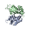
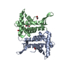
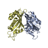
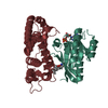
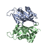
| タイトル | Crystal structure of ALBA2-DNA complex | ||||||
 要素 要素 |
| ||||||
 キーワード キーワード | DNA BINDING PROTEIN/DNA / ALBA 2 / Archaea / DNA-Binding Protein / Structural Genomics / NPPSFA / National Project on Protein Structural and Functional Analyses / RIKEN Structural Genomics/Proteomics Initiative / RSGI / DNA Binding Protein-DNA complex | ||||||
| 機能・相同性 |  機能・相同性情報 機能・相同性情報chromosome condensation / chromosome / double-stranded DNA binding / RNA binding / cytoplasm 類似検索 - 分子機能 | ||||||
| 生物種 |   Aeropyrum pernix (古細菌) Aeropyrum pernix (古細菌)synthetic construct (人工物) | ||||||
| 手法 |  X線回折 / シンクロトロン / 分子置換 / 解像度: 2 Å X線回折 / シンクロトロン / 分子置換 / 解像度: 2 Å | ||||||
 データ登録者 データ登録者 | Kumarevel, T. / Tanaka, T. / RIKEN Structural Genomics/Proteomics Initiative (RSGI) | ||||||
 引用 引用 | ジャーナル: J.Biol.Chem. / 年: 2012 タイトル: Crystal structure of archaeal chromatin protein Alba2-double-stranded DNA complex from Aeropyrum pernix K1. 著者: Tanaka, T. / Padavattan, S. / Kumarevel, T. #1: ジャーナル: Proteins / 年: 2008 タイトル: Crystal structure of an archaeal specific DNA-binding protein (Ape10b2) from Aeropyrum pernix K1. 著者: Kumarevel, T. / Sakamoto, K. / Gopinath, S.C. / Shinkai, A. / Kumar, P.K. / Yokoyama, S. | ||||||
| 履歴 |
|
- 構造の表示
構造の表示
| 構造ビューア | 分子: MolmilJmol/JSmol |
|---|
- ダウンロードとリンク
-ダウンロード
| PDBx/mmCIF形式 | 3u6y.cif.gz | 61.7 KB | 表示 | PDBx/mmCIF形式 |
|---|---|---|---|---|
| PDB形式 | pdb3u6y.ent.gz | 43.6 KB | 表示 | PDB形式 |
| PDBx/mmJSON形式 | 3u6y.json.gz | ツリー表示 | PDBx/mmJSON形式 | |
| その他 |  その他のダウンロード その他のダウンロード |
-検証レポート
| アーカイブディレクトリ | https://data.pdbj.org/pub/pdb/validation_reports/u6/3u6yftp://data.pdbj.org/pub/pdb/validation_reports/u6/3u6y | HTTPS FTP |
|---|
-関連構造データ
| 関連構造データ |  2h9uS S: 精密化の開始モデル |
|---|---|
| 類似構造データ | |
| その他のデータベース |


-リンク
 PDBj
PDBj
- 集合体
集合体
| 登録構造単位 | 
| ||||||||
|---|---|---|---|---|---|---|---|---|---|
| 1 |
| ||||||||
| 単位格子 |
|
-要素
| #1: タンパク質 | 分子量: 11397.197 Da / 分子数: 2 / 由来タイプ: 組換発現 / 由来: (組換発現) Aeropyrum pernix (古細菌) / 株: K1 / 遺伝子: albA2, APE_1823 / プラスミド: PET21A / 発現宿主:  #2: DNA鎖 | | 分子量: 1481.000 Da / 分子数: 1 / 由来タイプ: 合成 / 由来: (合成) synthetic construct (人工物) #3: DNA鎖 | | 分子量: 1521.024 Da / 分子数: 1 / 由来タイプ: 合成 / 由来: (合成) synthetic construct (人工物) #4: 化合物 | ChemComp-PO4 / |   分子量: 94.971 Da / 分子数: 1 / 由来タイプ: 合成 / 式: PO4 分子量: 94.971 Da / 分子数: 1 / 由来タイプ: 合成 / 式: PO4#5: 水 | ChemComp-HOH / |  分子量: 18.015 Da / 分子数: 183 / 由来タイプ: 天然 / 式: H2O 分子量: 18.015 Da / 分子数: 183 / 由来タイプ: 天然 / 式: H2OHas protein modification | Y | |
|---|
-実験情報
-実験
| 実験 | 手法: X線回折 / 使用した結晶の数: 1 |
|---|
- 試料調製
試料調製
| 結晶 | マシュー密度: 2.13 Å3/Da / 溶媒含有率: 42.27 % |
|---|---|
| 結晶化 | 温度: 293 K / 手法: 蒸気拡散法, シッティングドロップ法 / pH: 4.6 詳細: 0.2M AMMONIUM DIHYDROGEN PHOSPHATE, PEG 3350, pH 4.6, VAPOR DIFFUSION, SITTING DROP, temperature 293K |
-データ収集
| 回折 | 平均測定温度: 100 K |
|---|---|
| 放射光源 | 由来: シンクロトロン / サイト: SPring-8  / ビームライン: BL26B1 / 波長: 1 / 波長: 1 Å / ビームライン: BL26B1 / 波長: 1 / 波長: 1 Å |
| 検出器 | タイプ: ADSC QUANTUM 4 / 検出器: CCD / 日付: 2009年3月3日 / 詳細: SI II CHANNEL |
| 放射 | モノクロメーター: SI II CHANNEL / プロトコル: SINGLE WAVELENGTH / 単色(M)・ラウエ(L): M / 散乱光タイプ: x-ray |
| 放射波長 | 波長: 1 Å / 相対比: 1 |
| 反射 | 解像度: 2→40 Å / Num. all: 15617 / Num. obs: 15617 / % possible obs: 99.9 % / Observed criterion σ(F): 3 / Observed criterion σ(I): 3 / 冗長度: 14.1 % / Biso Wilson estimate: 19.1 Å2 / Rmerge(I) obs: 0.069 |
| 反射 シェル | 解像度: 2→2.07 Å / 冗長度: 14.3 % / Rmerge(I) obs: 0.496 / % possible all: 100 |
- 解析
解析
| ソフトウェア |
| ||||||||||||||||||||||||||||||||||||
|---|---|---|---|---|---|---|---|---|---|---|---|---|---|---|---|---|---|---|---|---|---|---|---|---|---|---|---|---|---|---|---|---|---|---|---|---|---|
| 精密化 | 構造決定の手法: 分子置換 開始モデル: 2H9U 解像度: 2→19.93 Å / Rfactor Rfree error: 0.008 / Data cutoff high absF: 1188968 / Data cutoff low absF: 0 / Isotropic thermal model: RESTRAINED / 交差検証法: THROUGHOUT / σ(F): 0 / 立体化学のターゲット値: Engh & Huber
| ||||||||||||||||||||||||||||||||||||
| 溶媒の処理 | 溶媒モデル: FLAT MODEL / Bsol: 54.36 Å2 / ksol: 0.34 e/Å3 | ||||||||||||||||||||||||||||||||||||
| 原子変位パラメータ | Biso mean: 44.03 Å2
| ||||||||||||||||||||||||||||||||||||
| Refine analyze |
| ||||||||||||||||||||||||||||||||||||
| 精密化ステップ | サイクル: LAST / 解像度: 2→19.93 Å
| ||||||||||||||||||||||||||||||||||||
| 拘束条件 |
| ||||||||||||||||||||||||||||||||||||
| LS精密化 シェル | 解像度: 2→2.13 Å / Rfactor Rfree error: 0.022 / Total num. of bins used: 6
| ||||||||||||||||||||||||||||||||||||
| Xplor file |
|
