 Movie
Movie Controller

[English] 日本語
 Yorodumi
- PDB-3tu9: Crystal structure of rabbit muscle aldolase bound with 5-O-methyl... -
+ Open data

- Basic information
| Entry | Database: PDB / ID: 3tu9 | ||||||
|---|---|---|---|---|---|---|---|
| Title | Crystal structure of rabbit muscle aldolase bound with 5-O-methyl mannitol 1,6-phosphate | ||||||
 Components Components | Fructose-bisphosphate aldolase A | ||||||
 Keywords Keywords | LYASE/LYASE INHIBITOR / beta-barrel / mammalian aldolase / mannitol-bisphosphate / trypanosomal aldolase / inhibitor docking / LYASE-LYASE INHIBITOR complex | ||||||
| Function / homology |  Function and homology information Function and homology informationnegative regulation of Arp2/3 complex-mediated actin nucleation / fructose-bisphosphate aldolase / fructose-bisphosphate aldolase activity / M band / I band / glycolytic process / protein homotetramerization / positive regulation of cell migration Similarity search - Function | ||||||
| Biological species |  | ||||||
| Method |  X-RAY DIFFRACTION / SYNCHROTRON / MOLECULAR REPLACEMENT / Resolution: 2.09 Å X-RAY DIFFRACTION / SYNCHROTRON / MOLECULAR REPLACEMENT / Resolution: 2.09 Å | ||||||
 Authors Authors | Arthus-Cartier, G. / Sygusch, J. | ||||||
 Citation Citation | Journal: ACS Med Chem Lett / Year: 2011 Title: Mannitol Bis-phosphate Based Inhibitors of Fructose 1,6-Bisphosphate Aldolases. Authors: Mabiala-Bassiloua, C.G. / Arthus-Cartier, G. / Hannaert, V. / Therisod, H. / Sygusch, J. / Therisod, M. | ||||||
| History |
|
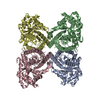
- Structure visualization
| Structure viewer | Molecule: MolmilJmol/JSmol |
|---|
- Downloads & links
-Download
| PDBx/mmCIF format | 3tu9.cif.gz | 307.8 KB | Display | PDBx/mmCIF format |
|---|---|---|---|---|
| PDB format | pdb3tu9.ent.gz | 248.8 KB | Display | PDB format |
| PDBx/mmJSON format | 3tu9.json.gz | Tree view | PDBx/mmJSON format | |
| Others |  Other downloads Other downloads |
-Validation report
| Arichive directory | https://data.pdbj.org/pub/pdb/validation_reports/tu/3tu9ftp://data.pdbj.org/pub/pdb/validation_reports/tu/3tu9 | HTTPS FTP |
|---|
-Related structure data
| Related structure data | 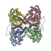 1zahS S: Starting model for refinement |
|---|---|
| Similar structure data |


-Links
 PDBj
PDBj
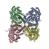
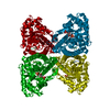
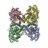
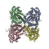
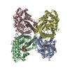
- Assembly
| Deposited unit | 
| ||||||||
|---|---|---|---|---|---|---|---|---|---|
| 1 |
| ||||||||
| Unit cell |
|
-Components
| #1: Protein | Mass: 39263.672 Da / Num. of mol.: 4 Source method: isolated from a genetically manipulated source Source: (gene. exp.)  #2: Sugar | ChemComp-5MM /   Type: D-saccharide / Mass: 356.158 Da / Num. of mol.: 4 Type: D-saccharide / Mass: 356.158 Da / Num. of mol.: 4Source method: isolated from a genetically manipulated source Formula: C7H18O12P2 #3: Water | ChemComp-HOH / |  Mass: 18.015 Da / Num. of mol.: 1420 / Source method: isolated from a natural source / Formula: H2O Mass: 18.015 Da / Num. of mol.: 1420 / Source method: isolated from a natural source / Formula: H2O |
|---|
-Experimental details
-Experiment
| Experiment | Method: X-RAY DIFFRACTION / Number of used crystals: 1 |
|---|
- Sample preparation
| Crystal | Density Matthews: 2.27 Å3/Da / Density % sol: 45.73 % |
|---|---|
| Crystal grow | Temperature: 298 K / Method: vapor diffusion, hanging drop / pH: 7.5 Details: 17.5% PEG4000, 0.1 M HEPES sodium, pH 7.5, VAPOR DIFFUSION, HANGING DROP, temperature 298K |
-Data collection
| Diffraction | Mean temperature: 100 K |
|---|---|
| Diffraction source | Source: SYNCHROTRON / Site: NSLS  / Beamline: X29A / Wavelength: 1.0809 Å / Beamline: X29A / Wavelength: 1.0809 Å |
| Detector | Type: ADSC QUANTUM 315 / Detector: CCD / Date: Nov 27, 2008 |
| Radiation | Monochromator: Rosenbaum-Rock double crystal sagittal focusing Protocol: SINGLE WAVELENGTH / Monochromatic (M) / Laue (L): M / Scattering type: x-ray |
| Radiation wavelength | Wavelength: 1.0809 Å / Relative weight: 1 |
| Reflection | Resolution: 2.09→36.973 Å / Num. all: 82812 / Num. obs: 82812 / % possible obs: 99.9 % / Observed criterion σ(F): 1 / Observed criterion σ(I): 1 / Redundancy: 3.8 % / Rmerge(I) obs: 0.11 / Net I/σ(I): 12.4 |
| Reflection shell | Resolution: 2.09→2.16 Å / Redundancy: 3.6 % / Rmerge(I) obs: 0.589 / Mean I/σ(I) obs: 2.2 / % possible all: 99.6 |
- Processing
| Software |
| |||||||||||||||||||||||||||||||||||||||||||||||||||||||||||||||||||||||||||||||||||||||||||||||||||||||||||||||||||||||||||||||||||||||||||||||||||||||||||||||||||||||||||||||||||||||||||||||||||||||||||||||||||||||||
|---|---|---|---|---|---|---|---|---|---|---|---|---|---|---|---|---|---|---|---|---|---|---|---|---|---|---|---|---|---|---|---|---|---|---|---|---|---|---|---|---|---|---|---|---|---|---|---|---|---|---|---|---|---|---|---|---|---|---|---|---|---|---|---|---|---|---|---|---|---|---|---|---|---|---|---|---|---|---|---|---|---|---|---|---|---|---|---|---|---|---|---|---|---|---|---|---|---|---|---|---|---|---|---|---|---|---|---|---|---|---|---|---|---|---|---|---|---|---|---|---|---|---|---|---|---|---|---|---|---|---|---|---|---|---|---|---|---|---|---|---|---|---|---|---|---|---|---|---|---|---|---|---|---|---|---|---|---|---|---|---|---|---|---|---|---|---|---|---|---|---|---|---|---|---|---|---|---|---|---|---|---|---|---|---|---|---|---|---|---|---|---|---|---|---|---|---|---|---|---|---|---|---|---|---|---|---|---|---|---|---|---|---|---|---|---|---|---|---|
| Refinement | Method to determine structure: MOLECULAR REPLACEMENT Starting model: PDB ENTRY 1ZAH Resolution: 2.09→36.973 Å / SU ML: 0.24 / σ(F): 0.08 / Phase error: 18.64 / Stereochemistry target values: ML
| |||||||||||||||||||||||||||||||||||||||||||||||||||||||||||||||||||||||||||||||||||||||||||||||||||||||||||||||||||||||||||||||||||||||||||||||||||||||||||||||||||||||||||||||||||||||||||||||||||||||||||||||||||||||||
| Solvent computation | Shrinkage radii: 0.9 Å / VDW probe radii: 1.11 Å / Solvent model: FLAT BULK SOLVENT MODEL / Bsol: 46.134 Å2 / ksol: 0.43 e/Å3 | |||||||||||||||||||||||||||||||||||||||||||||||||||||||||||||||||||||||||||||||||||||||||||||||||||||||||||||||||||||||||||||||||||||||||||||||||||||||||||||||||||||||||||||||||||||||||||||||||||||||||||||||||||||||||
| Displacement parameters |
| |||||||||||||||||||||||||||||||||||||||||||||||||||||||||||||||||||||||||||||||||||||||||||||||||||||||||||||||||||||||||||||||||||||||||||||||||||||||||||||||||||||||||||||||||||||||||||||||||||||||||||||||||||||||||
| Refinement step | Cycle: LAST / Resolution: 2.09→36.973 Å
| |||||||||||||||||||||||||||||||||||||||||||||||||||||||||||||||||||||||||||||||||||||||||||||||||||||||||||||||||||||||||||||||||||||||||||||||||||||||||||||||||||||||||||||||||||||||||||||||||||||||||||||||||||||||||
| Refine LS restraints |
| |||||||||||||||||||||||||||||||||||||||||||||||||||||||||||||||||||||||||||||||||||||||||||||||||||||||||||||||||||||||||||||||||||||||||||||||||||||||||||||||||||||||||||||||||||||||||||||||||||||||||||||||||||||||||
| LS refinement shell |
|
