[English] 日本語
Yorodumi
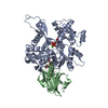
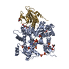
- PDB-3tu5: Actin complex with Gelsolin Segment 1 fused to Cobl segment -

+
Open data


ID or keywords:

Loading...

-
Basic information

Entry
Database: PDB / ID: 3tu5
TitleActin complex with Gelsolin Segment 1 fused to Cobl segment
Components
  • Actin, alpha skeletal muscle
  • Gelsolin,Protein cordon-bleu,Thymosin beta-4
KeywordsSTRUCTURAL PROTEIN/ACTIN-BINDING PROTEIN / Unusual hairpin conformation in the D-loop / STRUCTURAL PROTEIN-ACTIN-BINDING PROTEIN complex
Function / homology
Function and homology information


positive regulation of proton-transporting ATP synthase activity, rotational mechanism / somite specification / floor plate development / actin filament network formation / terminal web / striated muscle atrophy / regulation of establishment of T cell polarity / regulation of plasma membrane raft polarization / regulation of receptor clustering / renal protein absorption ...positive regulation of proton-transporting ATP synthase activity, rotational mechanism / somite specification / floor plate development / actin filament network formation / terminal web / striated muscle atrophy / regulation of establishment of T cell polarity / regulation of plasma membrane raft polarization / regulation of receptor clustering / renal protein absorption / positive regulation of keratinocyte apoptotic process / positive regulation of protein processing in phagocytic vesicle / embryonic axis specification / positive regulation of actin nucleation / phosphatidylinositol 3-kinase catalytic subunit binding / positive regulation of cysteine-type endopeptidase activity involved in apoptotic signaling pathway / actin cap / actin crosslink formation / sequestering of actin monomers / regulation of podosome assembly / notochord development / myosin II binding / negative regulation of viral entry into host cell / actin filament severing / actin filament capping / barbed-end actin filament capping / actin filament depolymerization / actin polymerization or depolymerization / digestive tract development / cell projection assembly / cardiac muscle cell contraction / positive regulation of endothelial cell chemotaxis / collateral sprouting in absence of injury / podosome / sarcoplasm / positive regulation of ruffle assembly / positive regulation of dendrite development / Sensory processing of sound by outer hair cells of the cochlea / relaxation of cardiac muscle / cytoskeletal motor activator activity / phagocytosis, engulfment / cortical actin cytoskeleton / tropomyosin binding / myosin heavy chain binding / mesenchyme migration / positive regulation of ATP biosynthetic process / troponin I binding / hepatocyte apoptotic process / actin filament bundle / filamentous actin / dendritic growth cone / actin filament bundle assembly / skeletal muscle thin filament assembly / striated muscle thin filament / skeletal muscle myofibril / cilium assembly / actin monomer binding / Caspase-mediated cleavage of cytoskeletal proteins / positive regulation of blood vessel endothelial cell migration / skeletal muscle fiber development / phagocytic vesicle / axonal growth cone / stress fiber / negative regulation of canonical NF-kappaB signal transduction / regulation of cell migration / titin binding / ruffle / tumor necrosis factor-mediated signaling pathway / phosphatidylinositol-4,5-bisphosphate binding / response to muscle stretch / actin filament polymerization / protein sequestering activity / platelet alpha granule lumen / filopodium / liver development / central nervous system development / actin filament organization / neural tube closure / actin filament / Hydrolases; Acting on acid anhydrides; Acting on acid anhydrides to facilitate cellular and subcellular movement / protein destabilization / negative regulation of inflammatory response / cellular response to type II interferon / calcium-dependent protein binding / actin filament binding / actin cytoskeleton / Platelet degranulation / lamellipodium / cell body / actin binding / cell cortex / actin cytoskeleton organization / blood microparticle / secretory granule lumen / ficolin-1-rich granule lumen / amyloid fibril formation / cytoskeleton / hydrolase activity / Amyloid fiber formation / axon
Similarity search - Function
Cordon-bleu, ubiquitin-like domain / Protein cordon-bleu-like / Cordon-bleu ubiquitin-like domain / Thymosin beta-4 family signature. / Beta-thymosin / Beta-thymosin superfamily / Thymosin beta-4 family / Thymosin beta actin-binding motif. / WH2 motif / Wiskott Aldrich syndrome homology region 2 ...Cordon-bleu, ubiquitin-like domain / Protein cordon-bleu-like / Cordon-bleu ubiquitin-like domain / Thymosin beta-4 family signature. / Beta-thymosin / Beta-thymosin superfamily / Thymosin beta-4 family / Thymosin beta actin-binding motif. / WH2 motif / Wiskott Aldrich syndrome homology region 2 / WH2 domain / WH2 domain profile. / Actin; Chain A, domain 2 / Actin; Chain A, domain 2 / Villin/Gelsolin / Gelsolin homology domain / Severin / Severin / Gelsolin-like domain / Gelsolin repeat / ADF-H/Gelsolin-like domain superfamily / ATPase, substrate binding domain, subdomain 4 / Actin; Chain A, domain 4 / ATPase, nucleotide binding domain / Actins signature 1. / Actin, conserved site / Actins signature 2. / Actin/actin-like conserved site / Actins and actin-related proteins signature. / Actin / Actin family / Actin / ATPase, nucleotide binding domain / Nucleotidyltransferase; domain 5 / Roll / Alpha-Beta Complex / 2-Layer Sandwich / 3-Layer(aba) Sandwich / Mainly Beta / Alpha Beta
Similarity search - Domain/homology
ADENOSINE-5'-TRIPHOSPHATE / Gelsolin / Thymosin beta-4 / Actin, alpha skeletal muscle / Protein cordon-bleu
Similarity search - Component
Biological speciesHomo sapiens (human)
Mus musculus (house mouse)
Oryctolagus cuniculus (rabbit)
MethodX-RAY DIFFRACTION / SYNCHROTRON / MOLECULAR REPLACEMENT / molecular replacement / Resolution: 3 Å
AuthorsKudryashov, D.S. / Sawaya, M.R. / Durer, Z.A.O.
CitationJournal: Biophys.J. / Year: 2012
Title: Structural States and dynamics of the d-loop in actin.
Authors: Durer, Z.A. / Kudryashov, D.S. / Sawaya, M.R. / Altenbach, C. / Hubbell, W. / Reisler, E.
History
DepositionSep 15, 2011Deposition site: RCSB / Processing site: RCSB
Revision 1.0Sep 26, 2012Provider: repository / Type: Initial release
Revision 1.1Oct 10, 2012Group: Database references
Revision 1.2Jun 7, 2017Group: Database references
Revision 1.3Feb 28, 2024Group: Data collection / Database references / Derived calculations
Category: chem_comp_atom / chem_comp_bond ...chem_comp_atom / chem_comp_bond / database_2 / pdbx_struct_conn_angle / struct_conn / struct_site
Item: _database_2.pdbx_DOI / _database_2.pdbx_database_accession ..._database_2.pdbx_DOI / _database_2.pdbx_database_accession / _pdbx_struct_conn_angle.ptnr1_auth_asym_id / _pdbx_struct_conn_angle.ptnr1_auth_comp_id / _pdbx_struct_conn_angle.ptnr1_auth_seq_id / _pdbx_struct_conn_angle.ptnr1_label_asym_id / _pdbx_struct_conn_angle.ptnr1_label_atom_id / _pdbx_struct_conn_angle.ptnr1_label_comp_id / _pdbx_struct_conn_angle.ptnr1_label_seq_id / _pdbx_struct_conn_angle.ptnr2_auth_asym_id / _pdbx_struct_conn_angle.ptnr2_auth_seq_id / _pdbx_struct_conn_angle.ptnr2_label_asym_id / _pdbx_struct_conn_angle.ptnr3_auth_asym_id / _pdbx_struct_conn_angle.ptnr3_auth_comp_id / _pdbx_struct_conn_angle.ptnr3_auth_seq_id / _pdbx_struct_conn_angle.ptnr3_label_asym_id / _pdbx_struct_conn_angle.ptnr3_label_atom_id / _pdbx_struct_conn_angle.ptnr3_label_comp_id / _pdbx_struct_conn_angle.ptnr3_label_seq_id / _pdbx_struct_conn_angle.value / _struct_conn.pdbx_dist_value / _struct_conn.ptnr1_auth_asym_id / _struct_conn.ptnr1_auth_comp_id / _struct_conn.ptnr1_auth_seq_id / _struct_conn.ptnr1_label_asym_id / _struct_conn.ptnr1_label_atom_id / _struct_conn.ptnr1_label_comp_id / _struct_conn.ptnr1_label_seq_id / _struct_conn.ptnr2_auth_asym_id / _struct_conn.ptnr2_auth_comp_id / _struct_conn.ptnr2_auth_seq_id / _struct_conn.ptnr2_label_asym_id / _struct_conn.ptnr2_label_atom_id / _struct_conn.ptnr2_label_comp_id / _struct_site.pdbx_auth_asym_id / _struct_site.pdbx_auth_comp_id / _struct_site.pdbx_auth_seq_id

-
Structure visualization

Structure viewerMolecule:
MolmilJmol/JSmol

Downloads & links

-
Assembly

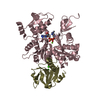
Deposited unit
A: Actin, alpha skeletal muscle
B: Gelsolin,Protein cordon-bleu,Thymosin beta-4
hetero molecules


Theoretical massNumber of molelcules
Total (without water)75,4147
Polymers74,5902
Non-polymers8245
Water1,17165
1


  • Idetical with deposited unit
  • defined by author&software
TypeNameSymmetry operationNumber
identity operation1_555x,y,z1
Buried area4480 Å2
ΔGint-67 kcal/mol
Surface area21980 Å2
MethodPISA
Unit cell
Length a, b, c (Å)71.184, 71.184, 222.244
Angle α, β, γ (deg.)90.000, 90.000, 120.000
Int Tables number154
Space group name H-MP3221

-
Components

-
Protein , 2 types, 2 molecules AB

#1: Protein Actin, alpha skeletal muscle / / Alpha-actin-1


Mass: 42096.953 Da / Num. of mol.: 1 / Source method: isolated from a natural source / Details: skeletal / Source: (natural) Oryctolagus cuniculus (rabbit) / References: UniProt: P68135
#2: Protein Gelsolin,Protein cordon-bleu,Thymosin beta-4 / AGEL / Actin-depolymerizing factor / ADF / Brevin / T beta-4 / Fx


Mass: 32493.480 Da / Num. of mol.: 1
Source method: isolated from a genetically manipulated source
Source: (gene. exp.) Homo sapiens (human), (gene. exp.) Mus musculus (house mouse)
Gene: GSN, Cobl, Kiaa0633, TMSB4X, TB4X, THYB4, TMSB4 / Plasmid: pCOLD-1 / Production host: Escherichia coli (E. coli) / Strain (production host): Rosetta-2 cells
References: UniProt: P06396, UniProt: Q5NBX1, UniProt: P62328

-
Non-polymers , 4 types, 70 molecules

#3: Chemical ChemComp-CA / CALCIUM ION


Mass: 40.078 Da / Num. of mol.: 2 / Source method: obtained synthetically / Formula: Ca
#4: Chemical ChemComp-ATP / ADENOSINE-5'-TRIPHOSPHATE / Adenosine triphosphate


Mass: 507.181 Da / Num. of mol.: 1 / Source method: obtained synthetically / Formula: C10H16N5O13P3 / Comment: ATP, energy-carrying molecule*YM
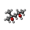
#5: Chemical ChemComp-MPD / (4S)-2-METHYL-2,4-PENTANEDIOL / 2-Methyl-2,4-pentanediol


Mass: 118.174 Da / Num. of mol.: 2 / Source method: obtained synthetically / Formula: C6H14O2 / Comment: precipitant*YM
#6: Water ChemComp-HOH / water / Water


Mass: 18.015 Da / Num. of mol.: 65 / Source method: isolated from a natural source / Formula: H2O

-
Experimental details

-
Experiment

ExperimentMethod: X-RAY DIFFRACTION / Number of used crystals: 1

-
Sample preparation

CrystalDensity Matthews: 2.18 Å3/Da / Density % sol: 43.56 %
Crystal growTemperature: 298 K / Method: vapor diffusion, hanging drop / pH: 9
Details: 10% (w/v) polyethyleneglycol 20000, 2% (v/v) dioxane, and 0.1M bicine, pH 9.0, VAPOR DIFFUSION, HANGING DROP, temperature 298K

-
Data collection

DiffractionMean temperature: 100 K
Diffraction sourceSource: SYNCHROTRON / Site: APS / Beamline: 24-ID-C / Wavelength: 0.9794 Å
DetectorType: ADSC QUANTUM 315 / Detector: CCD / Date: Jul 22, 2010
RadiationMonochromator: Si (111) / Protocol: SINGLE WAVELENGTH / Monochromatic (M) / Laue (L): M / Scattering type: x-ray
Radiation wavelengthWavelength: 0.9794 Å / Relative weight: 1
ReflectionResolution: 3→118 Å / Num. all: 13348 / Num. obs: 13348 / % possible obs: 96.3 % / Observed criterion σ(I): -3 / Redundancy: 7 % / Biso Wilson estimate: 67.4 Å2 / Rmerge(I) obs: 0.089 / Χ2: 1.031 / Net I/σ(I): 11
Reflection shell
Resolution (Å)Redundancy (%)Rmerge(I) obsNum. unique allΧ2Diffraction-ID% possible all
3-3.117.20.49313321.121198.1
3.11-3.237.20.33913161.155197.9
3.23-3.387.10.22713171.15197.6
3.38-3.567.20.1613331.1197.6
3.56-3.787.10.12313121.001197.2
3.78-4.077.10.09713120.902196.8
4.07-4.487.10.08813460.994196.3
4.48-5.1370.07813320.992195.7
5.13-6.466.80.06113471.004194.7
6.46-1186.70.0414010.887191.8

-
Phasing

PhasingMethod: molecular replacement
Phasing MRModel details: Phaser MODE: MR_AUTO
Highest resolutionLowest resolution
Rotation3.7 Å33.8 Å
Translation3.7 Å33.8 Å

-
Processing

Software
NameVersionClassificationNB
DENZOdata reduction
SCALEPACKdata scaling
PHASER2.1.4phasing
BUSTER-TNTrefinement
PDB_EXTRACT3.1data extraction
ADSCQuantumdata collection
BUSTER2.8.0refinement
RefinementMethod to determine structure: MOLECULAR REPLACEMENT / Resolution: 3→25.42 Å / Cor.coef. Fo:Fc: 0.9505 / Cor.coef. Fo:Fc free: 0.9378 / Occupancy max: 1 / Occupancy min: 1 / Cross valid method: THROUGHOUT / σ(F): 0 / Stereochemistry target values: Engh & Huber
RfactorNum. reflection% reflectionSelection details
Rfree0.2045 668 5.02 %RANDOM
Rwork0.1652 ---
all0.1672 13348 --
obs0.1672 13297 --
Displacement parametersBiso max: 200.15 Å2 / Biso mean: 74.2852 Å2 / Biso min: 25.74 Å2
Baniso -1Baniso -2Baniso -3
1--4.2979 Å20 Å20 Å2
2---4.2979 Å20 Å2
3---8.5958 Å2
Refine analyzeLuzzati coordinate error obs: 0.392 Å
Refinement stepCycle: LAST / Resolution: 3→25.42 Å
ProteinNucleic acidLigandSolventTotal
Num. atoms3943 0 49 65 4057
Refine LS restraints
Refine-IDTypeNumberRestraint functionWeightDev ideal
X-RAY DIFFRACTIONt_dihedral_angle_d1420SINUSOIDAL2
X-RAY DIFFRACTIONt_trig_c_planes103HARMONIC2
X-RAY DIFFRACTIONt_gen_planes585HARMONIC5
X-RAY DIFFRACTIONt_it4077HARMONIC20
X-RAY DIFFRACTIONt_nbd0SEMIHARMONIC5
X-RAY DIFFRACTIONt_improper_torsion4HARMONIC0
X-RAY DIFFRACTIONt_pseud_angle
X-RAY DIFFRACTIONt_chiral_improper_torsion529SEMIHARMONIC5
X-RAY DIFFRACTIONt_sum_occupancies
X-RAY DIFFRACTIONt_utility_distance
X-RAY DIFFRACTIONt_utility_angle
X-RAY DIFFRACTIONt_utility_torsion
X-RAY DIFFRACTIONt_ideal_dist_contact4673SEMIHARMONIC4
X-RAY DIFFRACTIONt_bond_d4077HARMONIC20.01
X-RAY DIFFRACTIONt_angle_deg5529HARMONIC21.11
X-RAY DIFFRACTIONt_omega_torsion3.07
X-RAY DIFFRACTIONt_other_torsion22.43
LS refinement shellResolution: 3→3.24 Å / Total num. of bins used: 7
RfactorNum. reflection% reflection
Rfree0.2872 140 5.12 %
Rwork0.19 2597 -
all0.1948 2737 -
Refinement TLS params.

Method: refined / Refine-ID: X-RAY DIFFRACTION

IDL112)L122)L132)L222)L232)L332)S11 (Å °)S12 (Å °)S13 (Å °)S21 (Å °)S22 (Å °)S23 (Å °)S31 (Å °)S32 (Å °)S33 (Å °)T112)T122)T132)T222)T232)T332)Origin x (Å)Origin y (Å)Origin z (Å)
11.15440.1752-0.47182.8935-0.74294.7536-0.14130.0167-0.04580.47570.19150.0249-0.0442-0.611-0.0501-0.1291-0.0834-0.01-0.2648-0.0183-0.27516.3444-8.070922.1341
21.72971.0545-0.50713.3524-0.33523.4015-0.19950.0896-0.1088-0.39470.0887-0.32050.25850.02170.11090.0696-0.18260.1555-0.1761-0.0903-0.089630.7241-17.7104-3.9211
Refinement TLS group
IDRefine-IDRefine TLS-IDSelection detailsAuth asym-IDAuth seq-ID
1X-RAY DIFFRACTION1{ A|* }A4 - 377
2X-RAY DIFFRACTION2{ B|* }B1 - 132

+
About Yorodumi

-
News

-
Feb 9, 2022. New format data for meta-information of EMDB entries

New format data for meta-information of EMDB entries

  • Version 3 of the EMDB header file is now the official format.
  • The previous official version 1.9 will be removed from the archive.

Related info.:EMDB header

External links:wwPDB to switch to version 3 of the EMDB data model

-
Aug 12, 2020. Covid-19 info

Covid-19 info

URL: https://pdbj.org/emnavi/covid19.php

New page: Covid-19 featured information page in EM Navigator.

Related info.:Covid-19 info / Mar 5, 2020. Novel coronavirus structure data

+
Mar 5, 2020. Novel coronavirus structure data

Novel coronavirus structure data

Related info.:Yorodumi Speices / Aug 12, 2020. Covid-19 info

External links:COVID-19 featured content - PDBj / Molecule of the Month (242):Coronavirus Proteases

+
Jan 31, 2019. EMDB accession codes are about to change! (news from PDBe EMDB page)

EMDB accession codes are about to change! (news from PDBe EMDB page)

  • The allocation of 4 digits for EMDB accession codes will soon come to an end. Whilst these codes will remain in use, new EMDB accession codes will include an additional digit and will expand incrementally as the available range of codes is exhausted. The current 4-digit format prefixed with “EMD-” (i.e. EMD-XXXX) will advance to a 5-digit format (i.e. EMD-XXXXX), and so on. It is currently estimated that the 4-digit codes will be depleted around Spring 2019, at which point the 5-digit format will come into force.
  • The EM Navigator/Yorodumi systems omit the EMD- prefix.

Related info.:Q: What is EMD? / ID/Accession-code notation in Yorodumi/EM Navigator

External links:EMDB Accession Codes are Changing Soon! / Contact to PDBj

+
Jul 12, 2017. Major update of PDB

Major update of PDB

  • wwPDB released updated PDB data conforming to the new PDBx/mmCIF dictionary.
  • This is a major update changing the version number from 4 to 5, and with Remediation, in which all the entries are updated.
  • In this update, many items about electron microscopy experimental information are reorganized (e.g. em_software).
  • Now, EM Navigator and Yorodumi are based on the updated data.

External links:wwPDB Remediation / Enriched Model Files Conforming to OneDep Data Standards Now Available in the PDB FTP Archive

-
Yorodumi

Thousand views of thousand structures

  • Yorodumi is a browser for structure data from EMDB, PDB, SASBDB, etc.
  • This page is also the successor to EM Navigator detail page, and also detail information page/front-end page for Omokage search.
  • The word "yorodu" (or yorozu) is an old Japanese word meaning "ten thousand". "mi" (miru) is to see.

Related info.:EMDB / PDB / SASBDB / Comparison of 3 databanks / Yorodumi Search / Aug 31, 2016. New EM Navigator & Yorodumi / Yorodumi Papers / Jmol/JSmol / Function and homology information / Changes in new EM Navigator and Yorodumi

Read more