 Movie
Movie Controller

[English] 日本語
 Yorodumi
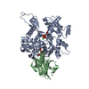
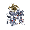
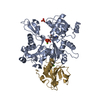
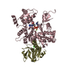
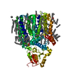
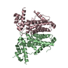
- PDB-4fww: Crystal structure of the Sema-PSI extracellular domains of human ... -
+ Open data

- Basic information
| Entry | Database: PDB / ID: 4fww | |||||||||
|---|---|---|---|---|---|---|---|---|---|---|
| Title | Crystal structure of the Sema-PSI extracellular domains of human RON receptor tyrosine kinase | |||||||||
 Components Components | Macrophage-stimulating protein receptor | |||||||||
 Keywords Keywords | TRANSFERASE / beta-propeller / cysteine-knot / receptor tyrosine kinase / macrophage stimulating protein / N-glycosylation / extracellular | |||||||||
| Function / homology |  Function and homology information Function and homology informationSignaling by MST1 / macrophage colony-stimulating factor receptor activity / vacuole / positive regulation of MAP kinase activity / single fertilization / phagocytosis / stress fiber / transmembrane receptor protein tyrosine kinase activity / cell surface receptor protein tyrosine kinase signaling pathway / defense response ...Signaling by MST1 / macrophage colony-stimulating factor receptor activity / vacuole / positive regulation of MAP kinase activity / single fertilization / phagocytosis / stress fiber / transmembrane receptor protein tyrosine kinase activity / cell surface receptor protein tyrosine kinase signaling pathway / defense response / receptor protein-tyrosine kinase / response to virus / nervous system development / cell migration / positive regulation of phosphatidylinositol 3-kinase/protein kinase B signal transduction / signaling receptor complex / innate immune response / positive regulation of cell population proliferation / enzyme binding / cell surface / signal transduction / ATP binding / plasma membrane Similarity search - Function | |||||||||
| Biological species |  Homo sapiens (human) Homo sapiens (human) | |||||||||
| Method |  X-RAY DIFFRACTION / SYNCHROTRON / MOLECULAR REPLACEMENT / Resolution: 1.85 Å X-RAY DIFFRACTION / SYNCHROTRON / MOLECULAR REPLACEMENT / Resolution: 1.85 Å | |||||||||
 Authors Authors | Chao, K.L. / Herzberg, O. | |||||||||
 Citation Citation | Journal: Plos One / Year: 2012 Title: Crystal Structure of the Sema-PSI Extracellular Domain of Human RON Receptor Tyrosine Kinase. Authors: Chao, K.L. / Tsai, I.W. / Chen, C. / Herzberg, O. | |||||||||
| History |
|
- Structure visualization
| Structure viewer | Molecule: MolmilJmol/JSmol |
|---|
- Downloads & links
-Download
| PDBx/mmCIF format | 4fww.cif.gz | 223.9 KB | Display | PDBx/mmCIF format |
|---|---|---|---|---|
| PDB format | pdb4fww.ent.gz | 176.9 KB | Display | PDB format |
| PDBx/mmJSON format | 4fww.json.gz | Tree view | PDBx/mmJSON format | |
| Others |  Other downloads Other downloads |
-Validation report
| Arichive directory | https://data.pdbj.org/pub/pdb/validation_reports/fw/4fwwftp://data.pdbj.org/pub/pdb/validation_reports/fw/4fww | HTTPS FTP |
|---|
-Related structure data
| Related structure data |  2uzxS S: Starting model for refinement |
|---|---|
| Similar structure data |

-Links
 PDBj
PDBj

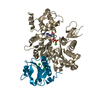
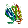
- Assembly
| Deposited unit | 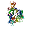
| ||||||||
|---|---|---|---|---|---|---|---|---|---|
| 1 |
| ||||||||
| Unit cell |
|
-Components
-Protein / Sugars , 2 types, 2 molecules A
| #1: Protein | Mass: 56132.543 Da / Num. of mol.: 1 Fragment: extracellular Sema and PSI domains (UNP Residues 42-568) Source method: isolated from a genetically manipulated source Details: The furin consensus cleavage sequence was mutated to a thrombin cleavage sequence. The recombinant protein contained the 115 amino acid residues IPT1 domain. However, proteolytic degradation ...Details: The furin consensus cleavage sequence was mutated to a thrombin cleavage sequence. The recombinant protein contained the 115 amino acid residues IPT1 domain. However, proteolytic degradation during crystallization removed ~75 amino acid residues of the domain and the remaining 40 residues are disordered and are not listed in the sequence record. Proteolytic degradation also removed N-terminal residues 23-41. These are not included in the sequence. Source: (gene. exp.) Homo sapiens (human) / Gene: MST1R, PTK8, RON / Plasmid: pMT/BiP-V5-HisA / Production host:  References: UniProt: Q04912, receptor protein-tyrosine kinase |
|---|---|
| #2: Polysaccharide | beta-D-mannopyranose-(1-3)-beta-D-mannopyranose-(1-4)-2-acetamido-2-deoxy-beta-D-glucopyranose-(1-4) ...beta-D-mannopyranose-(1-3)-beta-D-mannopyranose-(1-4)-2-acetamido-2-deoxy-beta-D-glucopyranose-(1-4)-2-acetamido-2-deoxy-beta-D-glucopyranose Source method: isolated from a genetically manipulated source |
-Non-polymers , 6 types, 374 molecules 


| #3: Chemical |  Mass: 96.063 Da / Num. of mol.: 2 / Source method: obtained synthetically / Formula: SO4 Mass: 96.063 Da / Num. of mol.: 2 / Source method: obtained synthetically / Formula: SO4#4: Chemical | ChemComp-ACT /  Mass: 59.044 Da / Num. of mol.: 5 / Source method: obtained synthetically / Formula: C2H3O2 Mass: 59.044 Da / Num. of mol.: 5 / Source method: obtained synthetically / Formula: C2H3O2#5: Chemical | ChemComp-GOL /  Mass: 92.094 Da / Num. of mol.: 4 / Source method: obtained synthetically / Formula: C3H8O3 Mass: 92.094 Da / Num. of mol.: 4 / Source method: obtained synthetically / Formula: C3H8O3#6: Chemical | ChemComp-EDO /  Mass: 62.068 Da / Num. of mol.: 4 / Source method: obtained synthetically / Formula: C2H6O2 Mass: 62.068 Da / Num. of mol.: 4 / Source method: obtained synthetically / Formula: C2H6O2#7: Chemical | ChemComp-PEG / |  Mass: 106.120 Da / Num. of mol.: 1 / Source method: obtained synthetically / Formula: C4H10O3 Mass: 106.120 Da / Num. of mol.: 1 / Source method: obtained synthetically / Formula: C4H10O3#8: Water | ChemComp-HOH / | Mass: 18.015 Da / Num. of mol.: 358 / Source method: isolated from a natural source / Formula: H2O |
|---|
-Details
| Has protein modification | Y |
|---|
-Experimental details
-Experiment
| Experiment | Method: X-RAY DIFFRACTION / Number of used crystals: 1 |
|---|
- Sample preparation
| Crystal | Density Matthews: 3.03 Å3/Da / Density % sol: 59.38 % |
|---|---|
| Crystal grow | Temperature: 298 K / Method: vapor diffusion, hanging drop / pH: 4.6 Details: 19% PEG 4000, 0.1 M Sodium Acetate, pH 4.6, 0.2 M Ammonium Sulfate, VAPOR DIFFUSION, HANGING DROP, temperature 298K |
-Data collection
| Diffraction | Mean temperature: 298 K |
|---|---|
| Diffraction source | Source: SYNCHROTRON / Site: APS  / Beamline: 23-ID-B / Wavelength: 1.0332 Å / Beamline: 23-ID-B / Wavelength: 1.0332 Å |
| Detector | Type: MARMOSAIC 300 mm CCD / Detector: CCD / Date: Feb 24, 2010 / Details: mirrors |
| Radiation | Monochromator: Si 111 Channel / Protocol: SINGLE WAVELENGTH / Monochromatic (M) / Laue (L): M / Scattering type: x-ray |
| Radiation wavelength | Wavelength: 1.0332 Å / Relative weight: 1 |
| Reflection | Resolution: 1.85→19.32 Å / Num. all: 57269 / Num. obs: 51510 / % possible obs: 90 % / Observed criterion σ(F): 0 / Observed criterion σ(I): -3 / Redundancy: 2.8 % / Rmerge(I) obs: 0.069 / Net I/σ(I): 10.46 |
| Reflection shell | Resolution: 1.85→1.9 Å / Redundancy: 2 % / Rmerge(I) obs: 0.44 / Mean I/σ(I) obs: 1.47 / % possible all: 63.7 |
- Processing
| Software |
| ||||||||||||||||||||||||||||||||||||||||||||||||||||||||||||||||||||||||||||||||||||||||||||||||||||||||||||||||||||||||||||||||||||||||||||||||||||||||||||||||||||||||||
|---|---|---|---|---|---|---|---|---|---|---|---|---|---|---|---|---|---|---|---|---|---|---|---|---|---|---|---|---|---|---|---|---|---|---|---|---|---|---|---|---|---|---|---|---|---|---|---|---|---|---|---|---|---|---|---|---|---|---|---|---|---|---|---|---|---|---|---|---|---|---|---|---|---|---|---|---|---|---|---|---|---|---|---|---|---|---|---|---|---|---|---|---|---|---|---|---|---|---|---|---|---|---|---|---|---|---|---|---|---|---|---|---|---|---|---|---|---|---|---|---|---|---|---|---|---|---|---|---|---|---|---|---|---|---|---|---|---|---|---|---|---|---|---|---|---|---|---|---|---|---|---|---|---|---|---|---|---|---|---|---|---|---|---|---|---|---|---|---|---|---|---|
| Refinement | Method to determine structure: MOLECULAR REPLACEMENT Starting model: PDB entry 2UZX chain B Resolution: 1.85→19.32 Å / Cor.coef. Fo:Fc: 0.962 / Cor.coef. Fo:Fc free: 0.943 / SU B: 3.857 / SU ML: 0.11 / Isotropic thermal model: Isotropic / Cross valid method: THROUGHOUT / ESU R: 0.137 / ESU R Free: 0.134 / Stereochemistry target values: MAXIMUM LIKELIHOOD / Details: HYDROGENS HAVE BEEN USED IF PRESENT IN THE INPUT
| ||||||||||||||||||||||||||||||||||||||||||||||||||||||||||||||||||||||||||||||||||||||||||||||||||||||||||||||||||||||||||||||||||||||||||||||||||||||||||||||||||||||||||
| Solvent computation | Ion probe radii: 0.8 Å / Shrinkage radii: 0.8 Å / VDW probe radii: 1.2 Å / Solvent model: MASK | ||||||||||||||||||||||||||||||||||||||||||||||||||||||||||||||||||||||||||||||||||||||||||||||||||||||||||||||||||||||||||||||||||||||||||||||||||||||||||||||||||||||||||
| Displacement parameters | Biso mean: 51.634 Å2
| ||||||||||||||||||||||||||||||||||||||||||||||||||||||||||||||||||||||||||||||||||||||||||||||||||||||||||||||||||||||||||||||||||||||||||||||||||||||||||||||||||||||||||
| Refinement step | Cycle: LAST / Resolution: 1.85→19.32 Å
| ||||||||||||||||||||||||||||||||||||||||||||||||||||||||||||||||||||||||||||||||||||||||||||||||||||||||||||||||||||||||||||||||||||||||||||||||||||||||||||||||||||||||||
| Refine LS restraints |
| ||||||||||||||||||||||||||||||||||||||||||||||||||||||||||||||||||||||||||||||||||||||||||||||||||||||||||||||||||||||||||||||||||||||||||||||||||||||||||||||||||||||||||
| LS refinement shell | Resolution: 1.85→1.898 Å / Total num. of bins used: 20
| ||||||||||||||||||||||||||||||||||||||||||||||||||||||||||||||||||||||||||||||||||||||||||||||||||||||||||||||||||||||||||||||||||||||||||||||||||||||||||||||||||||||||||
| Refinement TLS params. | Method: refined / Refine-ID: X-RAY DIFFRACTION
| ||||||||||||||||||||||||||||||||||||||||||||||||||||||||||||||||||||||||||||||||||||||||||||||||||||||||||||||||||||||||||||||||||||||||||||||||||||||||||||||||||||||||||
| Refinement TLS group |
|
