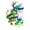
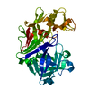
Entry Database : PDB / ID : 3tkmTitle Crystal structure PPAR delta binding GW0742 Peroxisome proliferator-activated receptor delta Keywords / / / Function / homology Function Domain/homology Component
/ / / / / / / / / / / / / / / / / / / / / / / / / / / / / / / / / / / / / / / / / / / / / / / / / / / / / / / / / / / / / / / / / / / / / / / / / / / / / / / / / / / / / / / / / / / / / / / / / / / / / / / / / / / / / / / / / / / / / Biological species Homo sapiens (human)Method / / / Resolution : 1.953 Å Authors Trivella, D.B.B. / Batista, F.H. / Polikarpov, I. Journal : Plos One / Year : 2012Title : Structural Insights into Human Peroxisome Proliferator Activated Receptor Delta (PPAR-Delta) Selective Ligand Binding.Authors : Batista, F.A. / Trivella, D.B. / Bernardes, A. / Gratieri, J. / Oliveira, P.S. / Figueira, A.C. / Webb, P. / Polikarpov, I. History Deposition Aug 27, 2011 Deposition site / Processing site Revision 1.0 Jul 4, 2012 Provider / Type Revision 1.1 Oct 21, 2020 Group / Database references / Derived calculationsCategory reflns / reflns_shell ... reflns / reflns_shell / struct_ref_seq_dif / struct_site Item _reflns.pdbx_Rmerge_I_obs / _reflns.pdbx_Rsym_value ... _reflns.pdbx_Rmerge_I_obs / _reflns.pdbx_Rsym_value / _reflns_shell.Rmerge_I_obs / _reflns_shell.number_unique_all / _reflns_shell.number_unique_obs / _struct_ref_seq_dif.details / _struct_site.pdbx_auth_asym_id / _struct_site.pdbx_auth_comp_id / _struct_site.pdbx_auth_seq_id Revision 1.2 Sep 13, 2023 Group / Database references / Refinement descriptionCategory chem_comp_atom / chem_comp_bond ... chem_comp_atom / chem_comp_bond / database_2 / pdbx_initial_refinement_model Item / _database_2.pdbx_database_accession
Show all Show less
 Movie
Movie Controller


 Open data
 Basic information
 Components
 Keywords
 Function and homology information
 Homo sapiens (human)
 X-RAY DIFFRACTION /
  Authors
 Citation
 Structure visualization
 Downloads & links
 Other downloads


 PDBj
PDBj
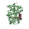
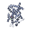
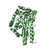
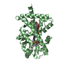
 Assembly


 Mass: 471.488 Da / Num. of mol.: 1 / Source method: obtained synthetically / Formula: C21H17F4NO3S2 / Comment: agonist*YM
 Mass: 92.094 Da / Num. of mol.: 1 / Source method: obtained synthetically / Formula: C3H8O3
 Mass: 18.015 Da / Num. of mol.: 158 / Source method: isolated from a natural source / Formula: H2O
 Sample preparation
 / Beamline: W01B-MX2 / Wavelength: 1.4589 Å
 Processing