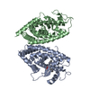
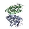
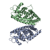
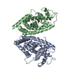
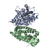
[English] 日本語
Yorodumi
- PDB-3cdp: Crystal structure of PPAR-gamma LBD complexed with a partial agon... -

+
Open data


ID or keywords:

Loading...

-
Basic information

Entry
Database: PDB / ID: 3cdp
TitleCrystal structure of PPAR-gamma LBD complexed with a partial agonist, analogue of clofibric acid
ComponentsPeroxisome proliferator-activated receptor gamma
KeywordsTRANSCRIPTION / bundle of alpha-helices and a small four-stranded beta-sheet / Activator / Diabetes mellitus / Disease mutation / DNA-binding / Metal-binding / Nucleus / Obesity / Phosphoprotein / Receptor / Transcription regulation / Zinc-finger
Function / homology
Function and homology information


prostaglandin receptor activity / negative regulation of receptor signaling pathway via STAT / MECP2 regulates transcription factors / negative regulation of vascular endothelial cell proliferation / negative regulation of extracellular matrix assembly / negative regulation of connective tissue replacement involved in inflammatory response wound healing / positive regulation of cholesterol transport / negative regulation of cellular response to transforming growth factor beta stimulus / : / arachidonate binding ...prostaglandin receptor activity / negative regulation of receptor signaling pathway via STAT / MECP2 regulates transcription factors / negative regulation of vascular endothelial cell proliferation / negative regulation of extracellular matrix assembly / negative regulation of connective tissue replacement involved in inflammatory response wound healing / positive regulation of cholesterol transport / negative regulation of cellular response to transforming growth factor beta stimulus / : / arachidonate binding / positive regulation of adiponectin secretion / negative regulation of cardiac muscle hypertrophy in response to stress / DNA binding domain binding / lipoprotein transport / positive regulation of vascular associated smooth muscle cell apoptotic process / WW domain binding / STAT family protein binding / positive regulation of fatty acid metabolic process / response to lipid / negative regulation of SMAD protein signal transduction / negative regulation of type II interferon-mediated signaling pathway / LBD domain binding / negative regulation of cholesterol storage / lipid homeostasis / E-box binding / alpha-actinin binding / negative regulation of vascular associated smooth muscle cell proliferation / R-SMAD binding / monocyte differentiation / negative regulation of blood vessel endothelial cell migration / white fat cell differentiation / negative regulation of macrophage derived foam cell differentiation / negative regulation of lipid storage / cellular response to low-density lipoprotein particle stimulus / negative regulation of BMP signaling pathway / positive regulation of cholesterol efflux / negative regulation of mitochondrial fission / cell fate commitment / negative regulation of osteoblast differentiation / positive regulation of fat cell differentiation / negative regulation of MAPK cascade / BMP signaling pathway / long-chain fatty acid transport / nuclear retinoid X receptor binding / Transcriptional regulation of brown and beige adipocyte differentiation by EBF2 / retinoic acid receptor signaling pathway / cell maturation / intracellular receptor signaling pathway / positive regulation of adipose tissue development / hormone-mediated signaling pathway / peroxisome proliferator activated receptor signaling pathway / epithelial cell differentiation / regulation of cellular response to insulin stimulus / response to nutrient / negative regulation of miRNA transcription / peptide binding / negative regulation of angiogenesis / transcription coregulator binding / positive regulation of apoptotic signaling pathway / Regulation of PTEN gene transcription / fatty acid metabolic process / placenta development / SUMOylation of intracellular receptors / negative regulation of smooth muscle cell proliferation / negative regulation of transforming growth factor beta receptor signaling pathway / mRNA transcription by RNA polymerase II / PPARA activates gene expression / regulation of circadian rhythm / Nuclear Receptor transcription pathway / Transcriptional regulation of white adipocyte differentiation / positive regulation of miRNA transcription / regulation of blood pressure / lipid metabolic process / DNA-binding transcription repressor activity, RNA polymerase II-specific / negative regulation of inflammatory response / RNA polymerase II transcription regulator complex / cellular response to insulin stimulus / nuclear receptor activity / rhythmic process / glucose homeostasis / MLL4 and MLL3 complexes regulate expression of PPARG target genes in adipogenesis and hepatic steatosis / DNA-binding transcription activator activity, RNA polymerase II-specific / double-stranded DNA binding / DNA-binding transcription factor binding / cellular response to hypoxia / sequence-specific DNA binding / nucleic acid binding / cell differentiation / DNA-binding transcription factor activity, RNA polymerase II-specific / receptor complex / transcription cis-regulatory region binding / RNA polymerase II cis-regulatory region sequence-specific DNA binding / DNA-binding transcription factor activity / innate immune response / negative regulation of gene expression / negative regulation of DNA-templated transcription / intracellular membrane-bounded organelle / chromatin binding / positive regulation of gene expression / regulation of transcription by RNA polymerase II
Similarity search - Function
Peroxisome proliferator-activated receptor gamma / Peroxisome proliferator-activated receptor gamma, N-terminal / PPAR gamma N-terminal region / Peroxisome proliferator-activated receptor / : / Retinoid X Receptor / Retinoid X Receptor / Nuclear hormone receptor / Nuclear hormones receptors DNA-binding region signature. / Zinc finger, nuclear hormone receptor-type ...Peroxisome proliferator-activated receptor gamma / Peroxisome proliferator-activated receptor gamma, N-terminal / PPAR gamma N-terminal region / Peroxisome proliferator-activated receptor / : / Retinoid X Receptor / Retinoid X Receptor / Nuclear hormone receptor / Nuclear hormones receptors DNA-binding region signature. / Zinc finger, nuclear hormone receptor-type / Double treble clef zinc finger, C4 type / Nuclear hormone receptors DNA-binding domain profile. / c4 zinc finger in nuclear hormone receptors / Nuclear hormone receptor, ligand-binding domain / Nuclear hormone receptor-like domain superfamily / Ligand-binding domain of nuclear hormone receptor / Nuclear receptor (NR) ligand-binding (LBD) domain profile. / Ligand binding domain of hormone receptors / Zinc finger, NHR/GATA-type / Orthogonal Bundle / Mainly Alpha
Similarity search - Domain/homology
(2S)-2-(4-chlorophenoxy)-3-phenylpropanoic acid / Peroxisome proliferator-activated receptor gamma
Similarity search - Component
Biological speciesHomo sapiens (human)
MethodX-RAY DIFFRACTION / SYNCHROTRON / FOURIER SYNTHESIS / Resolution: 2.8 Å
AuthorsPochetti, G. / Montanari, R. / Mazza, F.
Citation
Journal: Bioorg.Med.Chem. / Year: 2012
Title: Synthesis, biological evaluation and molecular investigation of fluorinated peroxisome proliferator-activated receptors alpha/gamma dual agonists
Authors: Fracchiolla, G. / Laghezza, A. / Piemontese, L. / Parente, M. / Lavecchia, A. / Pochetti, G. / Montanari, R. / Di Giovanni, C. / Carbonara, G. / Tortorella, P. / Novellino, E. / Loiodice, F.
#1: Journal: Proc.Natl.Acad.Sci.Usa / Year: 2001
Title: Structural determinants of ligand binding selectivity between the peroxisome proliferator-activated receptors
Authors: Xu, H.E. / Lambert, M.H. / Montana, V.G. / Plunket, K.D. / Moore, L.B. / Collins, J.L. / Oplinger, J.A. / Kliewer, S.A. / Gampe, R.T. / McKee, D.D. / Moore, J.T. / Willson, T.M.
History
DepositionFeb 27, 2008Deposition site: RCSB / Processing site: PDBJ
Revision 1.0Jan 13, 2009Provider: repository / Type: Initial release
Revision 1.1Jul 13, 2011Group: Version format compliance
Revision 1.2Dec 25, 2013Group: Database references
Revision 1.3Mar 13, 2024Group: Data collection / Database references / Derived calculations
Category: chem_comp_atom / chem_comp_bond ...chem_comp_atom / chem_comp_bond / database_2 / struct_ref_seq_dif / struct_site
Item: _database_2.pdbx_DOI / _database_2.pdbx_database_accession ..._database_2.pdbx_DOI / _database_2.pdbx_database_accession / _struct_ref_seq_dif.details / _struct_site.pdbx_auth_asym_id / _struct_site.pdbx_auth_comp_id / _struct_site.pdbx_auth_seq_id

-
Structure visualization

Structure viewerMolecule:
MolmilJmol/JSmol

Downloads & links

-
Assembly

Deposited unit
A: Peroxisome proliferator-activated receptor gamma
B: Peroxisome proliferator-activated receptor gamma
hetero molecules


Theoretical massNumber of molelcules
Total (without water)65,3383
Polymers65,0612
Non-polymers2771
Water1,17165
1


  • Idetical with deposited unit
  • defined by author
TypeNameSymmetry operationNumber
identity operation1_555x,y,z1
2
A: Peroxisome proliferator-activated receptor gamma
hetero molecules


Theoretical massNumber of molelcules
Total (without water)32,8072
Polymers32,5311
Non-polymers2771
Water181
TypeNameSymmetry operationNumber
identity operation1_555x,y,z1
MethodPISA
3
B: Peroxisome proliferator-activated receptor gamma


Theoretical massNumber of molelcules
Total (without water)32,5311
Polymers32,5311
Non-polymers00
Water181
TypeNameSymmetry operationNumber
identity operation1_555x,y,z1
MethodPISA
Unit cell
Length a, b, c (Å)93.210, 61.650, 118.360
Angle α, β, γ (deg.)90.00, 103.01, 90.00
Int Tables number5
Space group name H-MC121

-
Components

#1: Protein Peroxisome proliferator-activated receptor gamma / PPAR-gamma / Nuclear receptor subfamily 1 group C member 3


Mass: 32530.652 Da / Num. of mol.: 2 / Fragment: ligand binding domain (LBD), UNP residues 223-504
Source method: isolated from a genetically manipulated source
Source: (gene. exp.) Homo sapiens (human) / Plasmid: pET28 / Production host: Escherichia coli (E. coli) / Strain (production host): BL21 DE3 / References: UniProt: P37231
#2: Chemical ChemComp-YRG / (2S)-2-(4-chlorophenoxy)-3-phenylpropanoic acid


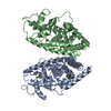
Mass: 276.715 Da / Num. of mol.: 1 / Source method: obtained synthetically / Formula: C15H13ClO3
#3: Water ChemComp-HOH / water


Mass: 18.015 Da / Num. of mol.: 65 / Source method: isolated from a natural source / Formula: H2O

-
Experimental details

-
Experiment

ExperimentMethod: X-RAY DIFFRACTION / Number of used crystals: 1

-
Sample preparation

CrystalDensity Matthews: 2.55 Å3/Da / Density % sol: 51.7 %
Crystal growTemperature: 293 K / Method: vapor diffusion, sitting drop / pH: 8
Details: 0.8M NaCitrate, 0.15mM Tris, pH 8.0, VAPOR DIFFUSION, SITTING DROP, temperature 293K

-
Data collection

DiffractionMean temperature: 100 K
Diffraction sourceSource: SYNCHROTRON / Site: ESRF / Beamline: ID14-1 / Wavelength: 0.934 Å
DetectorType: ADSC QUANTUM 210 / Detector: CCD / Date: Nov 10, 2007 / Details: bent mirrors
RadiationMonochromator: diamond 111 / Protocol: SINGLE WAVELENGTH / Monochromatic (M) / Laue (L): M / Scattering type: x-ray
Radiation wavelengthWavelength: 0.934 Å / Relative weight: 1
ReflectionResolution: 2.8→20 Å / Num. all: 16156 / Num. obs: 16156 / % possible obs: 99.1 % / Observed criterion σ(F): 0 / Observed criterion σ(I): 0 / Redundancy: 3.6 % / Biso Wilson estimate: 58 Å2 / Rmerge(I) obs: 0.104 / Net I/σ(I): 6.6
Reflection shellResolution: 2.8→2.95 Å / Redundancy: 3.5 % / Rmerge(I) obs: 0.469 / Mean I/σ(I) obs: 1.6 / Num. unique all: 2332 / % possible all: 99.1

-
Processing

Software
NameVersionClassification
ADSCQuantumdata collection
CNSrefinement
MOSFLMdata reduction
SCALAdata scaling
CNSphasing
RefinementMethod to determine structure: FOURIER SYNTHESIS / Resolution: 2.8→10 Å / σ(F): 0 / Stereochemistry target values: Engh & Huber
RfactorNum. reflectionSelection details
Rfree0.298 772 random
Rwork0.236 --
all0.237 15796 -
obs0.237 15796 -
Refinement stepCycle: LAST / Resolution: 2.8→10 Å
ProteinNucleic acidLigandSolventTotal
Num. atoms4332 0 19 65 4416
Refine LS restraints
Refine-IDTypeDev ideal
X-RAY DIFFRACTIONc_bond_d0.008
X-RAY DIFFRACTIONc_angle_d1.25

+
About Yorodumi

-
News

-
Feb 9, 2022. New format data for meta-information of EMDB entries

New format data for meta-information of EMDB entries

  • Version 3 of the EMDB header file is now the official format.
  • The previous official version 1.9 will be removed from the archive.

Related info.:EMDB header

External links:wwPDB to switch to version 3 of the EMDB data model

-
Aug 12, 2020. Covid-19 info

Covid-19 info

URL: https://pdbj.org/emnavi/covid19.php

New page: Covid-19 featured information page in EM Navigator.

Related info.:Covid-19 info / Mar 5, 2020. Novel coronavirus structure data

+
Mar 5, 2020. Novel coronavirus structure data

Novel coronavirus structure data

Related info.:Yorodumi Speices / Aug 12, 2020. Covid-19 info

External links:COVID-19 featured content - PDBj / Molecule of the Month (242):Coronavirus Proteases

+
Jan 31, 2019. EMDB accession codes are about to change! (news from PDBe EMDB page)

EMDB accession codes are about to change! (news from PDBe EMDB page)

  • The allocation of 4 digits for EMDB accession codes will soon come to an end. Whilst these codes will remain in use, new EMDB accession codes will include an additional digit and will expand incrementally as the available range of codes is exhausted. The current 4-digit format prefixed with “EMD-” (i.e. EMD-XXXX) will advance to a 5-digit format (i.e. EMD-XXXXX), and so on. It is currently estimated that the 4-digit codes will be depleted around Spring 2019, at which point the 5-digit format will come into force.
  • The EM Navigator/Yorodumi systems omit the EMD- prefix.

Related info.:Q: What is EMD? / ID/Accession-code notation in Yorodumi/EM Navigator

External links:EMDB Accession Codes are Changing Soon! / Contact to PDBj

+
Jul 12, 2017. Major update of PDB

Major update of PDB

  • wwPDB released updated PDB data conforming to the new PDBx/mmCIF dictionary.
  • This is a major update changing the version number from 4 to 5, and with Remediation, in which all the entries are updated.
  • In this update, many items about electron microscopy experimental information are reorganized (e.g. em_software).
  • Now, EM Navigator and Yorodumi are based on the updated data.

External links:wwPDB Remediation / Enriched Model Files Conforming to OneDep Data Standards Now Available in the PDB FTP Archive

-
Yorodumi

Thousand views of thousand structures

  • Yorodumi is a browser for structure data from EMDB, PDB, SASBDB, etc.
  • This page is also the successor to EM Navigator detail page, and also detail information page/front-end page for Omokage search.
  • The word "yorodu" (or yorozu) is an old Japanese word meaning "ten thousand". "mi" (miru) is to see.

Related info.:EMDB / PDB / SASBDB / Comparison of 3 databanks / Yorodumi Search / Aug 31, 2016. New EM Navigator & Yorodumi / Yorodumi Papers / Jmol/JSmol / Function and homology information / Changes in new EM Navigator and Yorodumi

Read more