 Movie
Movie Controller


+ Open data

- Basic information
| Entry | Database: PDB / ID: 3thw | ||||||
|---|---|---|---|---|---|---|---|
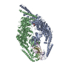
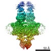
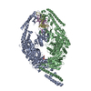
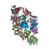
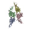
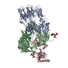
| Title | Human MutSbeta complexed with an IDL of 4 bases (Loop4) and ADP | ||||||
 Components Components |
| ||||||
 Keywords Keywords | DNA BINDING PROTEIN/DNA / ABC family ATPase / mismatch recognition / mismatched unpaired IDL DNA / DNA BINDING PROTEIN-DNA complex | ||||||
| Function / homology |  Function and homology information Function and homology informationsomatic recombination of immunoglobulin genes involved in immune response / MutSbeta complex / Defective Mismatch Repair Associated With MSH3 / MutSalpha complex / Defective Mismatch Repair Associated With MSH6 / Defective Mismatch Repair Associated With MSH2 / somatic recombination of immunoglobulin gene segments / guanine/thymine mispair binding / B cell mediated immunity / maintenance of DNA repeat elements ...somatic recombination of immunoglobulin genes involved in immune response / MutSbeta complex / Defective Mismatch Repair Associated With MSH3 / MutSalpha complex / Defective Mismatch Repair Associated With MSH6 / Defective Mismatch Repair Associated With MSH2 / somatic recombination of immunoglobulin gene segments / guanine/thymine mispair binding / B cell mediated immunity / maintenance of DNA repeat elements / positive regulation of isotype switching to IgA isotypes / centromeric DNA binding / positive regulation of isotype switching to IgG isotypes / mismatched DNA binding / mitotic recombination / negative regulation of DNA recombination / isotype switching / Mismatch repair (MMR) directed by MSH2:MSH3 (MutSbeta) / Mismatch repair (MMR) directed by MSH2:MSH6 (MutSalpha) / DNA damage tolerance / oxidative phosphorylation / response to UV-B / mitotic intra-S DNA damage checkpoint signaling / ATP-dependent DNA damage sensor activity / germ cell development / intrinsic apoptotic signaling pathway in response to DNA damage by p53 class mediator / response to X-ray / ATP-dependent activity, acting on DNA / mismatch repair / somatic hypermutation of immunoglobulin genes / B cell differentiation / determination of adult lifespan / TP53 Regulates Transcription of DNA Repair Genes / male gonad development / enzyme activator activity / double-strand break repair / double-stranded DNA binding / in utero embryonic development / damaged DNA binding / negative regulation of neuron apoptotic process / chromosome, telomeric region / DNA repair / chromatin binding / enzyme binding / protein homodimerization activity / ATP hydrolysis activity / DNA binding / nucleoplasm / ATP binding / membrane / nucleus Similarity search - Function | ||||||
| Biological species |  Homo sapiens (human) Homo sapiens (human) | ||||||
| Method |  X-RAY DIFFRACTION / SYNCHROTRON / MOLECULAR REPLACEMENT / Resolution: 3.09 Å X-RAY DIFFRACTION / SYNCHROTRON / MOLECULAR REPLACEMENT / Resolution: 3.09 Å | ||||||
 Authors Authors | Yang, W. | ||||||
 Citation Citation | Journal: Nat.Struct.Mol.Biol. / Year: 2012 Title: Mechanism of mismatch recognition revealed by human MutSbeta bound to unpaired DNA loops Authors: Gupta, S. / Gellert, M. / Yang, W. | ||||||
| History |
|
- Structure visualization
| Structure viewer | Molecule: MolmilJmol/JSmol |
|---|
- Downloads & links
-Download
| PDBx/mmCIF format | 3thw.cif.gz | 757 KB | Display | PDBx/mmCIF format |
|---|---|---|---|---|
| PDB format | pdb3thw.ent.gz | 613 KB | Display | PDB format |
| PDBx/mmJSON format | 3thw.json.gz | Tree view | PDBx/mmJSON format | |
| Others |  Other downloads Other downloads |
-Validation report
| Arichive directory | https://data.pdbj.org/pub/pdb/validation_reports/th/3thwftp://data.pdbj.org/pub/pdb/validation_reports/th/3thw | HTTPS FTP |
|---|
-Related structure data
| Related structure data |  3thxC 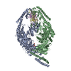 3thyC  3thzC  2o8bS C: citing same article ( S: Starting model for refinement |
|---|---|
| Similar structure data |


-Links
 PDBj
PDBj


- Assembly
| Deposited unit | 
| ||||||||
|---|---|---|---|---|---|---|---|---|---|
| 1 |
| ||||||||
| Unit cell |
|
-Components
| #1: Protein | Mass: 104861.875 Da / Num. of mol.: 1 Source method: isolated from a genetically manipulated source Source: (gene. exp.) Homo sapiens (human) / Gene: MSH2 / Cell line (production host): Hi5 / Production host:   Spodoptera frugiperda (fall armyworm) / References: UniProt: P43246 Spodoptera frugiperda (fall armyworm) / References: UniProt: P43246 |
|---|---|
| #2: Protein | Mass: 104289.664 Da / Num. of mol.: 1 / Fragment: UNP residues 219- 1134 Source method: isolated from a genetically manipulated source Source: (gene. exp.) Homo sapiens (human) / Gene: DUC1, DUG, MSH3 / Cell line (production host): Hi5 / Production host: Spodoptera frugiperda (fall armyworm) / References: GenBank: 119616268, UniProt: P20585*PLUS |
| #3: DNA chain | Mass: 16327.484 Da / Num. of mol.: 1 / Source method: obtained synthetically |
| #4: Chemical | ChemComp-ADP /   Mass: 427.201 Da / Num. of mol.: 1 / Source method: obtained synthetically / Formula: C10H15N5O10P2 / Comment: ADP, energy-carrying molecule*YM Mass: 427.201 Da / Num. of mol.: 1 / Source method: obtained synthetically / Formula: C10H15N5O10P2 / Comment: ADP, energy-carrying molecule*YM |
| #5: Water | ChemComp-HOH /  Mass: 18.015 Da / Num. of mol.: 4 / Source method: isolated from a natural source / Formula: H2O Mass: 18.015 Da / Num. of mol.: 4 / Source method: isolated from a natural source / Formula: H2O |
-Experimental details
-Experiment
| Experiment | Method: X-RAY DIFFRACTION / Number of used crystals: 1 |
|---|
- Sample preparation
| Crystal | Density Matthews: 2.45 Å3/Da / Density % sol: 49.87 % |
|---|---|
| Crystal grow | Temperature: 277 K / Method: vapor diffusion, hanging drop Details: 0.2M Ammonium Acetate, 20-25% PEG3350 (w/v) and 0.1M MES pH 6.5-7.5, VAPOR DIFFUSION, HANGING DROP, temperature 277K PH range: 6.5-7.5 |
-Data collection
| Diffraction | Mean temperature: 100 K |
|---|---|
| Diffraction source | Source: SYNCHROTRON / Site: APS  / Beamline: 22-BM / Wavelength: 1 Å / Beamline: 22-BM / Wavelength: 1 Å |
| Detector | Type: MARMOSAIC 225 mm CCD / Detector: CCD / Date: Dec 19, 2008 / Details: mirror |
| Radiation | Monochromator: Si 111 CHANNEL / Protocol: SINGLE WAVELENGTH / Monochromatic (M) / Laue (L): M / Scattering type: x-ray |
| Radiation wavelength | Wavelength: 1 Å / Relative weight: 1 |
| Reflection | Resolution: 3.09→33.2 Å / Num. obs: 41282 / % possible obs: 99 % / Observed criterion σ(F): 1 / Observed criterion σ(I): 1 / Redundancy: 2.4 % / Rmerge(I) obs: 0.0996 / Net I/σ(I): 7.5 |
| Reflection shell | Resolution: 3.09→3.14 Å / Redundancy: 2.4 % / Rmerge(I) obs: 0.554 / Mean I/σ(I) obs: 1.1 / Num. unique all: 1966 / % possible all: 98.5 |
- Processing
| Software |
| |||||||||||||||||||||||||||||||||||||||||||||||||||||||||||||||||||||||||||||
|---|---|---|---|---|---|---|---|---|---|---|---|---|---|---|---|---|---|---|---|---|---|---|---|---|---|---|---|---|---|---|---|---|---|---|---|---|---|---|---|---|---|---|---|---|---|---|---|---|---|---|---|---|---|---|---|---|---|---|---|---|---|---|---|---|---|---|---|---|---|---|---|---|---|---|---|---|---|---|
| Refinement | Method to determine structure: MOLECULAR REPLACEMENT Starting model: PDB ENTRY 2O8B Resolution: 3.09→32.53 Å / SU ML: 0.44 / σ(F): 0 / Phase error: 29.16 / Stereochemistry target values: ML
| |||||||||||||||||||||||||||||||||||||||||||||||||||||||||||||||||||||||||||||
| Solvent computation | Shrinkage radii: 0.83 Å / VDW probe radii: 1.1 Å / Solvent model: FLAT BULK SOLVENT MODEL / Bsol: 30.878 Å2 / ksol: 0.279 e/Å3 | |||||||||||||||||||||||||||||||||||||||||||||||||||||||||||||||||||||||||||||
| Displacement parameters |
| |||||||||||||||||||||||||||||||||||||||||||||||||||||||||||||||||||||||||||||
| Refinement step | Cycle: LAST / Resolution: 3.09→32.53 Å
| |||||||||||||||||||||||||||||||||||||||||||||||||||||||||||||||||||||||||||||
| Refine LS restraints |
| |||||||||||||||||||||||||||||||||||||||||||||||||||||||||||||||||||||||||||||
| LS refinement shell |
|
