 Movie
Movie Controller

[English] 日本語
 Yorodumi
- PDB-3tg9: The crystal structure of penicillin binding protein from Bacillus... -
+ Open data

- Basic information
| Entry | Database: PDB / ID: 3tg9 | ||||||
|---|---|---|---|---|---|---|---|
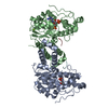
| Title | The crystal structure of penicillin binding protein from Bacillus halodurans | ||||||
 Components Components | Penicillin-binding protein | ||||||
 Keywords Keywords | penicillin binding protein / Structural Genomics / PSI-Biology / New York Structural Genomics Research Consortium / NYSGRC | ||||||
| Function / homology |  Function and homology information Function and homology information | ||||||
| Biological species |  Bacillus halodurans (bacteria) Bacillus halodurans (bacteria) | ||||||
| Method |  X-RAY DIFFRACTION / SYNCHROTRON / MOLECULAR REPLACEMENT / Resolution: 2.2 Å X-RAY DIFFRACTION / SYNCHROTRON / MOLECULAR REPLACEMENT / Resolution: 2.2 Å | ||||||
 Authors Authors | Zhang, Z. / Satyanarayana, L. / Chamala, S. / Evans, B. / Foti, R. / Gizzi, A. / Hillerich, B. / Kar, A. / LaFleur, J. / Seidel, R. ...Zhang, Z. / Satyanarayana, L. / Chamala, S. / Evans, B. / Foti, R. / Gizzi, A. / Hillerich, B. / Kar, A. / LaFleur, J. / Seidel, R. / Villigas, G. / Zencheck, W. / Almo, S.C. / Swaminathan, S. / New York Structural Genomics Research Consortium (NYSGRC) | ||||||
 Citation Citation | Journal: To be Published Title: The crystal structure of penicillin binding protein from Bacillus halodurans Authors: Zhang, Z. / Satyanarayana, L. / Almo, S.C. / Swaminathan, S. | ||||||
| History |
|
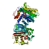
- Structure visualization
| Structure viewer | Molecule: MolmilJmol/JSmol |
|---|
- Downloads & links
-Download
| PDBx/mmCIF format | 3tg9.cif.gz | 143.7 KB | Display | PDBx/mmCIF format |
|---|---|---|---|---|
| PDB format | pdb3tg9.ent.gz | 111.5 KB | Display | PDB format |
| PDBx/mmJSON format | 3tg9.json.gz | Tree view | PDBx/mmJSON format | |
| Others |  Other downloads Other downloads |
-Validation report
| Summary document | 3tg9_validation.pdf.gz | 439.5 KB | Display | wwPDB validaton report |
|---|---|---|---|---|
| Full document | 3tg9_full_validation.pdf.gz | 447.2 KB | Display | |
| Data in XML | 3tg9_validation.xml.gz | 27 KB | Display | |
| Data in CIF | 3tg9_validation.cif.gz | 38.3 KB | Display | |
| Arichive directory | https://data.pdbj.org/pub/pdb/validation_reports/tg/3tg9ftp://data.pdbj.org/pub/pdb/validation_reports/tg/3tg9 | HTTPS FTP |
-Related structure data
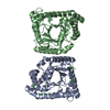
| Related structure data | 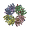 2qmiS S: Starting model for refinement |
|---|---|
| Similar structure data | |
| Other databases |


-Links
 PDBj
PDBj
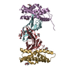
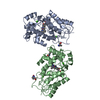
- Assembly
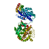
| Deposited unit | 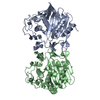
| ||||||||
|---|---|---|---|---|---|---|---|---|---|
| 1 |
| ||||||||
| Unit cell |
|
-Components
| #1: Protein | Mass: 40384.547 Da / Num. of mol.: 2 Source method: isolated from a genetically manipulated source Source: (gene. exp.) Bacillus halodurans (bacteria) / Strain: C-125 / Gene: BH2267 / Plasmid: pET / Production host: #2: Water | ChemComp-HOH / |  Mass: 18.015 Da / Num. of mol.: 267 / Source method: isolated from a natural source / Formula: H2O Mass: 18.015 Da / Num. of mol.: 267 / Source method: isolated from a natural source / Formula: H2O |
|---|
-Experimental details
-Experiment
| Experiment | Method: X-RAY DIFFRACTION / Number of used crystals: 1 |
|---|
- Sample preparation
| Crystal | Density Matthews: 2.36 Å3/Da / Density % sol: 47.86 % |
|---|---|
| Crystal grow | Temperature: 293 K / Method: vapor diffusion, sitting drop / pH: 5.5 Details: 0.2 M Ammonium sulfate, 0.1 M BIS-TRIS pH 5.5, 25% w/v Polyethylene glycol 3,350, 0.8% 1,4- butanediol, VAPOR DIFFUSION, SITTING DROP, temperature 293K |
-Data collection
| Diffraction | Mean temperature: 100 K |
|---|---|
| Diffraction source | Source: SYNCHROTRON / Site: NSLS  / Beamline: X29A / Wavelength: 0.9792 Å / Beamline: X29A / Wavelength: 0.9792 Å |
| Detector | Type: ADSC QUANTUM 315 / Detector: CCD / Date: Feb 7, 2011 / Details: mirrors |
| Radiation | Monochromator: Si 111 CHANNEL / Protocol: SINGLE WAVELENGTH / Monochromatic (M) / Laue (L): M / Scattering type: x-ray |
| Radiation wavelength | Wavelength: 0.9792 Å / Relative weight: 1 |
| Reflection | Resolution: 2.2→50 Å / Num. obs: 38211 / % possible obs: 99.9 % / Observed criterion σ(F): 0 / Observed criterion σ(I): 0 / Redundancy: 4.9 % / Rmerge(I) obs: 0.067 / Net I/σ(I): 20.4 |
| Reflection shell | Resolution: 2.2→2.28 Å / Redundancy: 4.7 % / Rmerge(I) obs: 0.248 / Mean I/σ(I) obs: 5.5 / Num. unique all: 3781 / % possible all: 99.2 |
- Processing
| Software |
| ||||||||||||||||||||||||||||||||||||||||||||||||||||||||||||||||||||||||||||||||||||||||||||||||||
|---|---|---|---|---|---|---|---|---|---|---|---|---|---|---|---|---|---|---|---|---|---|---|---|---|---|---|---|---|---|---|---|---|---|---|---|---|---|---|---|---|---|---|---|---|---|---|---|---|---|---|---|---|---|---|---|---|---|---|---|---|---|---|---|---|---|---|---|---|---|---|---|---|---|---|---|---|---|---|---|---|---|---|---|---|---|---|---|---|---|---|---|---|---|---|---|---|---|---|---|
| Refinement | Method to determine structure: MOLECULAR REPLACEMENT Starting model: PDB entry 2QMI Resolution: 2.2→49.999 Å / SU ML: 0.29 / Cross valid method: THROUGHOUT / σ(F): 0.19 / Stereochemistry target values: Engh & Huber
| ||||||||||||||||||||||||||||||||||||||||||||||||||||||||||||||||||||||||||||||||||||||||||||||||||
| Solvent computation | Shrinkage radii: 0.9 Å / VDW probe radii: 1.11 Å / Solvent model: FLAT BULK SOLVENT MODEL / Bsol: 49.334 Å2 / ksol: 0.378 e/Å3 | ||||||||||||||||||||||||||||||||||||||||||||||||||||||||||||||||||||||||||||||||||||||||||||||||||
| Displacement parameters |
| ||||||||||||||||||||||||||||||||||||||||||||||||||||||||||||||||||||||||||||||||||||||||||||||||||
| Refinement step | Cycle: LAST / Resolution: 2.2→49.999 Å
| ||||||||||||||||||||||||||||||||||||||||||||||||||||||||||||||||||||||||||||||||||||||||||||||||||
| Refine LS restraints |
| ||||||||||||||||||||||||||||||||||||||||||||||||||||||||||||||||||||||||||||||||||||||||||||||||||
| LS refinement shell |
|
