 Movie
Movie Controller

[English] 日本語
 Yorodumi
- PDB-2qmi: Structure of the octameric penicillin-binding protein homologue f... -
+ Open data

- Basic information
| Entry | Database: PDB / ID: 2qmi | ||||||
|---|---|---|---|---|---|---|---|
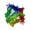
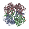
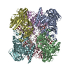
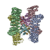
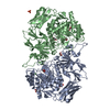
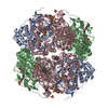
| Title | Structure of the octameric penicillin-binding protein homologue from Pyrococcus abyssi | ||||||
 Components Components | Pbp related beta-lactamase | ||||||
 Keywords Keywords | HYDROLASE / PAB87 / OCTAMER / LU-HPDO3A / PBP / ARCHAEA | ||||||
| Function / homology |  Function and homology information Function and homology informationPab87 octamerisation domain / Pab87 octamerisation domain / Pab87 octamerisation domain superfamily / Pab87 octamerisation domain / : / Beta-lactamase-related / Beta-lactamase / Beta-lactamase / DD-peptidase/beta-lactamase superfamily / Lipocalin ...Pab87 octamerisation domain / Pab87 octamerisation domain / Pab87 octamerisation domain superfamily / Pab87 octamerisation domain / : / Beta-lactamase-related / Beta-lactamase / Beta-lactamase / DD-peptidase/beta-lactamase superfamily / Lipocalin / Beta-lactamase/transpeptidase-like / Beta Barrel / 3-Layer(aba) Sandwich / Mainly Beta / Alpha Beta Similarity search - Domain/homology | ||||||
| Biological species |   Pyrococcus abyssi (archaea) Pyrococcus abyssi (archaea) | ||||||
| Method |  X-RAY DIFFRACTION / SYNCHROTRON / SAD / Resolution: 2.2 Å X-RAY DIFFRACTION / SYNCHROTRON / SAD / Resolution: 2.2 Å | ||||||
 Authors Authors | Delfosse, V. / Girard, E. / Moulinier, L. / Schultz, P. / Mayer, C. | ||||||
 Citation Citation | Journal: Plos One / Year: 2009 Title: Structure of the archaeal pab87 peptidase reveals a novel self-compartmentalizing protease family Authors: Delfosse, V. / Girard, E. / Birck, C. / Delmarcelle, M. / Delarue, M. / Poch, O. / Schultz, P. / Mayer, C. | ||||||
| History |
|
- Structure visualization
| Structure viewer | Molecule: MolmilJmol/JSmol |
|---|
- Downloads & links
-Download
| PDBx/mmCIF format | 2qmi.cif.gz | 735.7 KB | Display | PDBx/mmCIF format |
|---|---|---|---|---|
| PDB format | pdb2qmi.ent.gz | 606.4 KB | Display | PDB format |
| PDBx/mmJSON format | 2qmi.json.gz | Tree view | PDBx/mmJSON format | |
| Others |  Other downloads Other downloads |
-Validation report
| Arichive directory | https://data.pdbj.org/pub/pdb/validation_reports/qm/2qmiftp://data.pdbj.org/pub/pdb/validation_reports/qm/2qmi | HTTPS FTP |
|---|
-Related structure data
| Similar structure data |
|---|
-Links
 PDBj
PDBj

- Assembly
| Deposited unit | 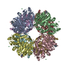
| ||||||||
|---|---|---|---|---|---|---|---|---|---|
| 1 |
| ||||||||
| Unit cell |
|
-Components
| #1: Protein | Mass: 50503.371 Da / Num. of mol.: 8 Source method: isolated from a genetically manipulated source Source: (gene. exp.) Pyrococcus abyssi (archaea) / Strain: GE5 / Gene: pbp / Plasmid: pET29a / Production host:  #2: Chemical | ChemComp-LU /   Mass: 174.967 Da / Num. of mol.: 17 / Source method: obtained synthetically / Formula: Lu Mass: 174.967 Da / Num. of mol.: 17 / Source method: obtained synthetically / Formula: Lu#3: Chemical | ChemComp-DO3 / 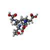  Mass: 404.459 Da / Num. of mol.: 4 / Source method: obtained synthetically / Formula: C17H32N4O7 Mass: 404.459 Da / Num. of mol.: 4 / Source method: obtained synthetically / Formula: C17H32N4O7#4: Water | ChemComp-HOH / |  Mass: 18.015 Da / Num. of mol.: 2368 / Source method: isolated from a natural source / Formula: H2O Mass: 18.015 Da / Num. of mol.: 2368 / Source method: isolated from a natural source / Formula: H2O |
|---|
-Experimental details
-Experiment
| Experiment | Method: X-RAY DIFFRACTION / Number of used crystals: 1 |
|---|
- Sample preparation
| Crystal | Density Matthews: 2.6 Å3/Da / Density % sol: 52.7 % |
|---|---|
| Crystal grow | Temperature: 291 K / Method: vapor diffusion, hanging drop / pH: 4.6 Details: 0.02M calcium chloride dihydrate, 0.1M sodium acetate trihydrate, 30% MPD, pH 4.6, VAPOR DIFFUSION, HANGING DROP, temperature 291K |
-Data collection
| Diffraction | Mean temperature: 100 K | ||||||||||||||||||||||||||||||||||||||||
|---|---|---|---|---|---|---|---|---|---|---|---|---|---|---|---|---|---|---|---|---|---|---|---|---|---|---|---|---|---|---|---|---|---|---|---|---|---|---|---|---|---|
| Diffraction source | Source: SYNCHROTRON / Site: ESRF  / Beamline: ID29 / Wavelength: 1.34029 Å / Beamline: ID29 / Wavelength: 1.34029 Å | ||||||||||||||||||||||||||||||||||||||||
| Detector | Type: ADSC QUANTUM 315 / Detector: CCD / Date: Feb 22, 2007 | ||||||||||||||||||||||||||||||||||||||||
| Radiation | Monochromator: Si 111 / Protocol: SINGLE WAVELENGTH / Monochromatic (M) / Laue (L): M / Scattering type: x-ray | ||||||||||||||||||||||||||||||||||||||||
| Radiation wavelength | Wavelength: 1.34029 Å / Relative weight: 1 | ||||||||||||||||||||||||||||||||||||||||
| Reflection | Av σ(I) over netI: 17.36 / Number: 1542549 / Rmerge(I) obs: 0.081 / D res high: 2.2 Å / Num. obs: 198621 / % possible obs: 95.9 | ||||||||||||||||||||||||||||||||||||||||
| Diffraction reflection shell |
| ||||||||||||||||||||||||||||||||||||||||
| Reflection | Resolution: 2.2→30 Å / Num. all: 198053 / Num. obs: 198053 / % possible obs: 95.7 % / Observed criterion σ(F): 0 / Observed criterion σ(I): 0 / Redundancy: 7.7 % / Biso Wilson estimate: 26.2 Å2 / Rsym value: 0.08 / Net I/σ(I): 17.4 | ||||||||||||||||||||||||||||||||||||||||
| Reflection shell | Resolution: 2.2→2.32 Å / Redundancy: 7.8 % / Mean I/σ(I) obs: 7.2 / Num. unique all: 28540 / Rsym value: 0.289 / % possible all: 93.8 |
- Processing
| Software |
| ||||||||||||||||||||||||||||
|---|---|---|---|---|---|---|---|---|---|---|---|---|---|---|---|---|---|---|---|---|---|---|---|---|---|---|---|---|---|
| Refinement | Method to determine structure: SAD / Resolution: 2.2→30 Å / Isotropic thermal model: Restrained / σ(F): 0 / σ(I): 0 / Stereochemistry target values: Engh & Huber
| ||||||||||||||||||||||||||||
| Solvent computation | Bsol: 37.475 Å2 | ||||||||||||||||||||||||||||
| Displacement parameters | Biso mean: 24.451 Å2
| ||||||||||||||||||||||||||||
| Refine analyze |
| ||||||||||||||||||||||||||||
| Refinement step | Cycle: LAST / Resolution: 2.2→30 Å
| ||||||||||||||||||||||||||||
| Refine LS restraints |
| ||||||||||||||||||||||||||||
| LS refinement shell | Resolution: 2.2→2.28 Å
| ||||||||||||||||||||||||||||
| Xplor file |
|
