| 登録情報 | データベース: PDB / ID: 3tdj
|
|---|
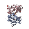
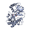
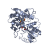
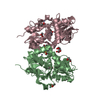
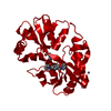
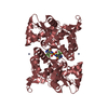
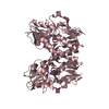
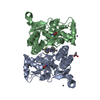
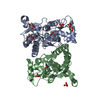
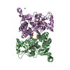
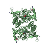
| タイトル | Crystal structure of the GluA2 ligand-binding domain (S1S2J-L483Y-N754S) in complex with glutamate and BPAM-97 at 1.95 A resolution |
|---|
 要素 要素 | Glutamate receptor 2 |
|---|
 キーワード キーワード | MEMBRANE PROTEIN / AMPA RECEPTOR LIGAND-BINDING DOMAIN / GLUA2 S1S2J-L483Y-N754S / BPAM-97 / ALLOSTERIC MODULATION |
|---|
| 機能・相同性 |  機能・相同性情報 機能・相同性情報
spine synapse / dendritic spine neck / dendritic spine head / cellular response to amine stimulus / Activation of AMPA receptors / perisynaptic space / ligand-gated monoatomic cation channel activity / AMPA glutamate receptor activity / Trafficking of GluR2-containing AMPA receptors / response to lithium ion ...spine synapse / dendritic spine neck / dendritic spine head / cellular response to amine stimulus / Activation of AMPA receptors / perisynaptic space / ligand-gated monoatomic cation channel activity / AMPA glutamate receptor activity / Trafficking of GluR2-containing AMPA receptors / response to lithium ion / kainate selective glutamate receptor activity / AMPA glutamate receptor complex / cellular response to glycine / extracellularly glutamate-gated ion channel activity / ionotropic glutamate receptor complex / immunoglobulin binding / asymmetric synapse / conditioned place preference / regulation of receptor recycling / glutamate receptor binding / Unblocking of NMDA receptors, glutamate binding and activation / positive regulation of synaptic transmission / regulation of synaptic transmission, glutamatergic / response to fungicide / glutamate-gated receptor activity / cytoskeletal protein binding / regulation of long-term synaptic depression / extracellular ligand-gated monoatomic ion channel activity / cellular response to brain-derived neurotrophic factor stimulus / glutamate-gated calcium ion channel activity / presynaptic active zone membrane / somatodendritic compartment / ionotropic glutamate receptor binding / dendrite membrane / ligand-gated monoatomic ion channel activity involved in regulation of presynaptic membrane potential / dendrite cytoplasm / ionotropic glutamate receptor signaling pathway / synaptic membrane / SNARE binding / dendritic shaft / transmitter-gated monoatomic ion channel activity involved in regulation of postsynaptic membrane potential / synaptic transmission, glutamatergic / PDZ domain binding / protein tetramerization / establishment of protein localization / postsynaptic density membrane / modulation of chemical synaptic transmission / cerebral cortex development / receptor internalization / Schaffer collateral - CA1 synapse / terminal bouton / synaptic vesicle / synaptic vesicle membrane / presynapse / signaling receptor activity / amyloid-beta binding / growth cone / presynaptic membrane / scaffold protein binding / perikaryon / chemical synaptic transmission / dendritic spine / postsynaptic membrane / neuron projection / postsynaptic density / axon / external side of plasma membrane / neuronal cell body / synapse / dendrite / protein kinase binding / protein-containing complex binding / glutamatergic synapse / cell surface / endoplasmic reticulum / protein-containing complex / identical protein binding / membrane / plasma membrane類似検索 - 分子機能 Ionotropic glutamate receptor, metazoa / Ligated ion channel L-glutamate- and glycine-binding site / Ionotropic glutamate receptor, L-glutamate and glycine-binding domain / Ligated ion channel L-glutamate- and glycine-binding site / Ligand-gated ion channel / : / Ionotropic glutamate receptor / Eukaryotic homologues of bacterial periplasmic substrate binding proteins. / Receptor, ligand binding region / Receptor family ligand binding region ...Ionotropic glutamate receptor, metazoa / Ligated ion channel L-glutamate- and glycine-binding site / Ionotropic glutamate receptor, L-glutamate and glycine-binding domain / Ligated ion channel L-glutamate- and glycine-binding site / Ligand-gated ion channel / : / Ionotropic glutamate receptor / Eukaryotic homologues of bacterial periplasmic substrate binding proteins. / Receptor, ligand binding region / Receptor family ligand binding region / Periplasmic binding protein-like II / Periplasmic binding protein-like I / D-Maltodextrin-Binding Protein; domain 2 / 3-Layer(aba) Sandwich / Alpha Beta類似検索 - ドメイン・相同性 Chem-3TJ / GLUTAMIC ACID / Glutamate receptor 2類似検索 - 構成要素 |
|---|
| 生物種 |   Rattus norvegicus (ドブネズミ) Rattus norvegicus (ドブネズミ) |
|---|
| 手法 |  X線回折 / シンクロトロン / 分子置換 / 解像度: 1.95 Å X線回折 / シンクロトロン / 分子置換 / 解像度: 1.95 Å |
|---|
 データ登録者 データ登録者 | Krintel, C. / Frydenvang, K. / Gajhede, M. / Kastrup, J.S. |
|---|
 引用 引用 | ジャーナル: Biochem.J. / 年: 2012タイトル: Thermodynamics and structural analysis of positive allosteric modulation of the ionotropic glutamate receptor GluA2. 著者: Krintel, C. / Frydenvang, K. / Olsen, L. / Kristensen, M.T. / de Barrios, O. / Naur, P. / Francotte, P. / Pirotte, B. / Gajhede, M. / Kastrup, J.S. |
|---|
| 履歴 | | 登録 | 2011年8月11日 | 登録サイト: RCSB / 処理サイト: RCSB |
|---|
| 改定 1.0 | 2011年9月21日 | Provider: repository / タイプ: Initial release |
|---|
| 改定 1.1 | 2011年12月28日 | Group: Database references |
|---|
| 改定 1.2 | 2017年8月9日 | Group: Data collection / Refinement description / Source and taxonomy
カテゴリ: diffrn_source / entity_src_gen / software / Item: _diffrn_source.pdbx_synchrotron_site |
|---|
| 改定 1.3 | 2023年9月13日 | Group: Data collection / Database references ...Data collection / Database references / Derived calculations / Refinement description
カテゴリ: chem_comp_atom / chem_comp_bond ...chem_comp_atom / chem_comp_bond / database_2 / pdbx_initial_refinement_model / struct_ref_seq_dif / struct_site
Item: _database_2.pdbx_DOI / _database_2.pdbx_database_accession ..._database_2.pdbx_DOI / _database_2.pdbx_database_accession / _struct_ref_seq_dif.details / _struct_site.pdbx_auth_asym_id / _struct_site.pdbx_auth_comp_id / _struct_site.pdbx_auth_seq_id |
|---|
| 改定 1.4 | 2024年11月20日 | Group: Structure summary
カテゴリ: pdbx_entry_details / pdbx_modification_feature |
|---|
|
|---|
 ムービー
ムービー コントローラー


 データを開く
 基本情報
基本情報 構造の表示
構造の表示 ダウンロードとリンク
 その他のダウンロード
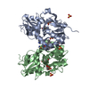


 PDBj
PDBj


 集合体
集合体


 タイプ: L-peptide linking / 分子量: 147.129 Da / 分子数: 2 / 由来タイプ: 合成 / 式: C5H9NO4
 分子量: 96.063 Da / 分子数: 3 / 由来タイプ: 合成 / 式: SO4
 分子量: 35.453 Da / 分子数: 5 / 由来タイプ: 合成 / 式: Cl
 分子量: 92.094 Da / 分子数: 11 / 由来タイプ: 合成 / 式: C3H8O3
 分子量: 230.259 Da / 分子数: 2 / 由来タイプ: 合成 / 式: C9H11FN2O2S
 試料調製
試料調製 / ビームライン: I911-5 / 波長: 0.908 Å
 解析
解析