 Movie
Movie Controller


+ Open data

- Basic information
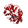
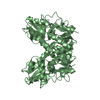
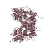
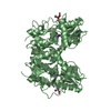
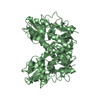
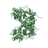
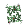
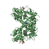
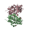
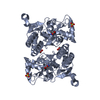
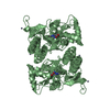
| Entry | Database: PDB / ID: 5h8s | ||||||
|---|---|---|---|---|---|---|---|
| Title | Structure of the human GluA2 LBD in complex with GNE3419 | ||||||
 Components Components | Glutamate receptor 2,Glutamate receptor 2 | ||||||
 Keywords Keywords | TRANSPORT PROTEIN / GluN1 / GluN2A / NMDA / receptor | ||||||
| Function / homology |  Function and homology information Function and homology informationActivation of AMPA receptors / Trafficking of GluR2-containing AMPA receptors / postsynaptic endocytic zone / ligand-gated monoatomic cation channel activity / AMPA glutamate receptor activity / Unblocking of NMDA receptors, glutamate binding and activation / AMPA glutamate receptor complex / Long-term potentiation / asymmetric synapse / MECP2 regulates neuronal receptors and channels ...Activation of AMPA receptors / Trafficking of GluR2-containing AMPA receptors / postsynaptic endocytic zone / ligand-gated monoatomic cation channel activity / AMPA glutamate receptor activity / Unblocking of NMDA receptors, glutamate binding and activation / AMPA glutamate receptor complex / Long-term potentiation / asymmetric synapse / MECP2 regulates neuronal receptors and channels / glutamate-gated receptor activity / excitatory synapse / ionotropic glutamate receptor signaling pathway / synaptic transmission, glutamatergic / transmitter-gated monoatomic ion channel activity involved in regulation of postsynaptic membrane potential / postsynaptic density membrane / modulation of chemical synaptic transmission / endocytic vesicle membrane / amyloid-beta binding / dendritic spine / postsynaptic density / postsynapse / external side of plasma membrane / neuronal cell body / dendrite / plasma membrane Similarity search - Function | ||||||
| Biological species |  Homo sapiens (human) Homo sapiens (human) | ||||||
| Method |  X-RAY DIFFRACTION / SYNCHROTRON / MOLECULAR REPLACEMENT / Resolution: 1.703 Å X-RAY DIFFRACTION / SYNCHROTRON / MOLECULAR REPLACEMENT / Resolution: 1.703 Å | ||||||
 Authors Authors | Wallweber, H.J.A. / Lupardus, P.J. | ||||||
 Citation Citation | Journal: Neuron / Year: 2016 Title: Positive Allosteric Modulators of GluN2A-Containing NMDARs with Distinct Modes of Action and Impacts on Circuit Function. Authors: Hackos, D.H. / Lupardus, P.J. / Grand, T. / Chen, Y. / Wang, T.M. / Reynen, P. / Gustafson, A. / Wallweber, H.J. / Volgraf, M. / Sellers, B.D. / Schwarz, J.B. / Paoletti, P. / Sheng, M. / Zhou, Q. / Hanson, J.E. | ||||||
| History |
|
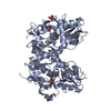
- Structure visualization
| Structure viewer | Molecule: MolmilJmol/JSmol |
|---|
- Downloads & links
-Download
| PDBx/mmCIF format | 5h8s.cif.gz | 183.4 KB | Display | PDBx/mmCIF format |
|---|---|---|---|---|
| PDB format | pdb5h8s.ent.gz | 145.2 KB | Display | PDB format |
| PDBx/mmJSON format | 5h8s.json.gz | Tree view | PDBx/mmJSON format | |
| Others |  Other downloads Other downloads |
-Validation report
| Arichive directory | https://data.pdbj.org/pub/pdb/validation_reports/h8/5h8sftp://data.pdbj.org/pub/pdb/validation_reports/h8/5h8s | HTTPS FTP |
|---|
-Related structure data
| Related structure data |  5h8fC  5h8hC  5h8nC  5h8qC  5kcjC  1ftjS C: citing same article ( S: Starting model for refinement |
|---|---|
| Similar structure data |


-Links
 PDBj
PDBj


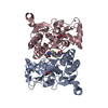
- Assembly
| Deposited unit | 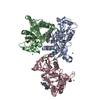
| ||||||||
|---|---|---|---|---|---|---|---|---|---|
| 1 | 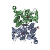
| ||||||||
| 2 | 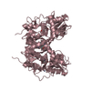
| ||||||||
| Unit cell |
|
-Components
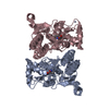
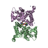
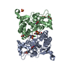
-Protein , 1 types, 3 molecules ACB
| #1: Protein | Mass: 29210.662 Da / Num. of mol.: 3 Fragment: UNP residues 413-527, GT linker, UNP residues 653-796 Source method: isolated from a genetically manipulated source Source: (gene. exp.) Homo sapiens (human) / Gene: GRIA2, GLUR2 / Production host:  |
|---|
-Non-polymers , 5 types, 678 molecules 


| #2: Chemical |  Type: L-peptide linking / Mass: 147.129 Da / Num. of mol.: 3 / Source method: obtained synthetically / Formula: C5H9NO4 Type: L-peptide linking / Mass: 147.129 Da / Num. of mol.: 3 / Source method: obtained synthetically / Formula: C5H9NO4#3: Chemical |  Mass: 300.379 Da / Num. of mol.: 2 / Source method: obtained synthetically / Formula: C15H16N4OS Mass: 300.379 Da / Num. of mol.: 2 / Source method: obtained synthetically / Formula: C15H16N4OS#4: Chemical | ChemComp-ZN /  Mass: 65.409 Da / Num. of mol.: 6 / Source method: obtained synthetically / Formula: Zn Mass: 65.409 Da / Num. of mol.: 6 / Source method: obtained synthetically / Formula: Zn#5: Chemical | ChemComp-CAC / |  Mass: 136.989 Da / Num. of mol.: 1 / Source method: obtained synthetically / Formula: C2H6AsO2 Mass: 136.989 Da / Num. of mol.: 1 / Source method: obtained synthetically / Formula: C2H6AsO2#6: Water | ChemComp-HOH / | Mass: 18.015 Da / Num. of mol.: 666 / Source method: isolated from a natural source / Formula: H2O |
|---|
-Details
| Has protein modification | Y |
|---|
-Experimental details
-Experiment
| Experiment | Method: X-RAY DIFFRACTION |
|---|
- Sample preparation
| Crystal | Density Matthews: 2.51 Å3/Da / Density % sol: 51 % |
|---|---|
| Crystal grow | Temperature: 277 K / Method: vapor diffusion, hanging drop / pH: 6.5 Details: 1:2 v/v protein to mother liquor (0.1 M HEPES, pH 7.0, 10-13% PEG8000, 2 mM calcium acetate) |
-Data collection
| Diffraction | Mean temperature: 100 K |
|---|---|
| Diffraction source | Source: SYNCHROTRON / Site: SSRL  / Beamline: BL11-1 / Wavelength: 0.979 Å / Beamline: BL11-1 / Wavelength: 0.979 Å |
| Detector | Type: DECTRIS PILATUS 6M / Detector: PIXEL / Date: Jan 5, 2012 |
| Radiation | Monochromator: Si(111) / Protocol: SINGLE WAVELENGTH / Monochromatic (M) / Laue (L): M / Scattering type: x-ray |
| Radiation wavelength | Wavelength: 0.979 Å / Relative weight: 1 |
| Reflection | Resolution: 1.7→50 Å / Num. obs: 96862 / % possible obs: 99.6 % / Redundancy: 6.5 % / Rmerge(I) obs: 0.075 / Net I/σ(I): 23.6 |
| Reflection shell | Resolution: 1.7→1.76 Å / Redundancy: 6.3 % / Rmerge(I) obs: 0.973 / Mean I/σ(I) obs: 2.2 / % possible all: 99.9 |
- Processing
| Software |
| |||||||||||||||||||||||||||||||||||||||||||||||||||||||||||||||||||||||||||||||||||||||||||||||||||||||||||||||||||||||||||||||||||||||||||||||||||||||||||||||||||||||||||||||||||||||||||||||||||||||||||||||||||||||||
|---|---|---|---|---|---|---|---|---|---|---|---|---|---|---|---|---|---|---|---|---|---|---|---|---|---|---|---|---|---|---|---|---|---|---|---|---|---|---|---|---|---|---|---|---|---|---|---|---|---|---|---|---|---|---|---|---|---|---|---|---|---|---|---|---|---|---|---|---|---|---|---|---|---|---|---|---|---|---|---|---|---|---|---|---|---|---|---|---|---|---|---|---|---|---|---|---|---|---|---|---|---|---|---|---|---|---|---|---|---|---|---|---|---|---|---|---|---|---|---|---|---|---|---|---|---|---|---|---|---|---|---|---|---|---|---|---|---|---|---|---|---|---|---|---|---|---|---|---|---|---|---|---|---|---|---|---|---|---|---|---|---|---|---|---|---|---|---|---|---|---|---|---|---|---|---|---|---|---|---|---|---|---|---|---|---|---|---|---|---|---|---|---|---|---|---|---|---|---|---|---|---|---|---|---|---|---|---|---|---|---|---|---|---|---|---|---|---|---|
| Refinement | Method to determine structure: MOLECULAR REPLACEMENT Starting model: PDB entry 1FTJ Resolution: 1.703→46.812 Å / SU ML: 0.19 / Cross valid method: FREE R-VALUE / σ(F): 1.34 / Phase error: 19.87 / Stereochemistry target values: ML
| |||||||||||||||||||||||||||||||||||||||||||||||||||||||||||||||||||||||||||||||||||||||||||||||||||||||||||||||||||||||||||||||||||||||||||||||||||||||||||||||||||||||||||||||||||||||||||||||||||||||||||||||||||||||||
| Solvent computation | Shrinkage radii: 0.9 Å / VDW probe radii: 1.11 Å / Solvent model: FLAT BULK SOLVENT MODEL | |||||||||||||||||||||||||||||||||||||||||||||||||||||||||||||||||||||||||||||||||||||||||||||||||||||||||||||||||||||||||||||||||||||||||||||||||||||||||||||||||||||||||||||||||||||||||||||||||||||||||||||||||||||||||
| Refinement step | Cycle: LAST / Resolution: 1.703→46.812 Å
| |||||||||||||||||||||||||||||||||||||||||||||||||||||||||||||||||||||||||||||||||||||||||||||||||||||||||||||||||||||||||||||||||||||||||||||||||||||||||||||||||||||||||||||||||||||||||||||||||||||||||||||||||||||||||
| Refine LS restraints |
| |||||||||||||||||||||||||||||||||||||||||||||||||||||||||||||||||||||||||||||||||||||||||||||||||||||||||||||||||||||||||||||||||||||||||||||||||||||||||||||||||||||||||||||||||||||||||||||||||||||||||||||||||||||||||
| LS refinement shell |
|
