 Movie
Movie Controller

[English] 日本語
 Yorodumi
- PDB-3td5: Crystal structure of OmpA-like domain from Acinetobacter baumanni... -
+ Open data

- Basic information
| Entry | Database: PDB / ID: 3td5 | |||||||||
|---|---|---|---|---|---|---|---|---|---|---|
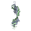
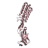
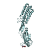
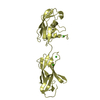
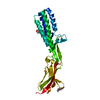
| Title | Crystal structure of OmpA-like domain from Acinetobacter baumannii in complex with L-Ala-gamma-D-Glu-m-DAP-D-Ala-D-Ala | |||||||||
 Components Components |
| |||||||||
 Keywords Keywords | MEMBRANE PROTEIN/PEPTIDE BINDING PROTEIN / OmpA-like fold / cell-wall attachment / peptidoglycan-binding / MEMBRANE PROTEIN-PEPTIDE BINDING PROTEIN complex | |||||||||
| Function / homology |  Function and homology information Function and homology informationporin activity / pore complex / host cell mitochondrion / monoatomic ion transport / cell outer membrane Similarity search - Function | |||||||||
| Biological species |  Acinetobacter baumannii (bacteria) Acinetobacter baumannii (bacteria) | |||||||||
| Method |  X-RAY DIFFRACTION / SYNCHROTRON / MOLECULAR REPLACEMENT / Resolution: 2 Å X-RAY DIFFRACTION / SYNCHROTRON / MOLECULAR REPLACEMENT / Resolution: 2 Å | |||||||||
 Authors Authors | Park, J.S. / Lee, W.C. / Song, J.H. / Kim, H.Y. | |||||||||
 Citation Citation | Journal: Faseb J. / Year: 2012 Title: Mechanism of anchoring of OmpA protein to the cell wall peptidoglycan of the gram-negative bacterial outer membrane Authors: Park, J.S. / Lee, W.C. / Yeo, K.J. / Ryu, K.S. / Kumarasiri, M. / Hesek, D. / Lee, M. / Mobashery, S. / Song, J.H. / Kim, S.I. / Lee, J.C. / Cheong, C. / Jeon, Y.H. / Kim, H.Y. | |||||||||
| History |
|
- Structure visualization
| Structure viewer | Molecule: MolmilJmol/JSmol |
|---|
- Downloads & links
-Download
| PDBx/mmCIF format | 3td5.cif.gz | 226.3 KB | Display | PDBx/mmCIF format |
|---|---|---|---|---|
| PDB format | pdb3td5.ent.gz | 182.9 KB | Display | PDB format |
| PDBx/mmJSON format | 3td5.json.gz | Tree view | PDBx/mmJSON format | |
| Others |  Other downloads Other downloads |
-Validation report
| Arichive directory | https://data.pdbj.org/pub/pdb/validation_reports/td/3td5ftp://data.pdbj.org/pub/pdb/validation_reports/td/3td5 | HTTPS FTP |
|---|
-Related structure data
| Related structure data |  3td3SC  3td4C S: Starting model for refinement C: citing same article ( |
|---|---|
| Similar structure data |


-Links
 PDBj
PDBj

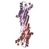
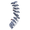
- Assembly
| Deposited unit | 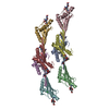
| ||||||||
|---|---|---|---|---|---|---|---|---|---|
| 1 | 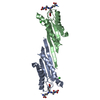
| ||||||||
| 2 | 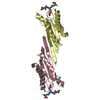
| ||||||||
| 3 | 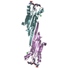
| ||||||||
| 4 | 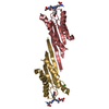
| ||||||||
| Unit cell |
|
-Components
| #1: Protein | Mass: 13879.479 Da / Num. of mol.: 8 / Fragment: C-TERMINAL DOMAIN Source method: isolated from a genetically manipulated source Source: (gene. exp.) Acinetobacter baumannii (bacteria) / Gene: omp38 / Production host: #2: Protein/peptide | Mass: 532.544 Da / Num. of mol.: 8 / Source method: obtained synthetically / Details: synthetic peptide #3: Chemical | ChemComp-CL /   Mass: 35.453 Da / Num. of mol.: 6 / Source method: obtained synthetically / Formula: Cl Mass: 35.453 Da / Num. of mol.: 6 / Source method: obtained synthetically / Formula: Cl#4: Water | ChemComp-HOH / |  Mass: 18.015 Da / Num. of mol.: 885 / Source method: isolated from a natural source / Formula: H2O Mass: 18.015 Da / Num. of mol.: 885 / Source method: isolated from a natural source / Formula: H2OCompound details | THE PEPTIDOGLYCAN IN CHAINS I, J, K, L, M, N, O, P CORRESPONDS TO MURAMYL PENTAPEPTIDE. HOWEVER, ...THE PEPTIDOGLY | |
|---|
-Experimental details
-Experiment
| Experiment | Method: X-RAY DIFFRACTION / Number of used crystals: 1 |
|---|
- Sample preparation
| Crystal | Density Matthews: 2.8 Å3/Da / Density % sol: 56.15 % |
|---|---|
| Crystal grow | Temperature: 293 K / Method: vapor diffusion, hanging drop / pH: 7.5 Details: 0.1M HEPES, 62.5%(v/v) 2-methyl-2,4-pentanediol , pH 7.5, VAPOR DIFFUSION, HANGING DROP, temperature 293K |
-Data collection
| Diffraction | Mean temperature: 100 K |
|---|---|
| Diffraction source | Source: SYNCHROTRON / Site: Photon Factory  / Beamline: BL-17A / Wavelength: 0.98 Å / Beamline: BL-17A / Wavelength: 0.98 Å |
| Detector | Type: ADSC QUANTUM 315r / Detector: CCD / Date: Jul 4, 2011 |
| Radiation | Monochromator: Si(111) crystals / Protocol: SINGLE WAVELENGTH / Monochromatic (M) / Laue (L): M / Scattering type: x-ray |
| Radiation wavelength | Wavelength: 0.98 Å / Relative weight: 1 |
| Reflection | Resolution: 2→81.3 Å / Num. obs: 78672 / % possible obs: 92.2 % / Observed criterion σ(F): 0 / Observed criterion σ(I): 0 |
| Reflection shell | Resolution: 2→2.11 Å / % possible all: 92.2 |
- Processing
| Software |
| ||||||||||||||||||||
|---|---|---|---|---|---|---|---|---|---|---|---|---|---|---|---|---|---|---|---|---|---|
| Refinement | Method to determine structure: MOLECULAR REPLACEMENT Starting model: 3TD3 Resolution: 2→81.3 Å / Cross valid method: THROUGHOUT / Stereochemistry target values: MAXIMUM LIKELIHOOD / Details: HYDROGENS HAVE BEEN ADDED IN THE RIDING POSITIONS
| ||||||||||||||||||||
| Refinement step | Cycle: LAST / Resolution: 2→81.3 Å
| ||||||||||||||||||||
| Refine LS restraints |
| ||||||||||||||||||||
| LS refinement shell | Resolution: 2→2.07 Å
|
