 Movie
Movie Controller

[English] 日本語
 Yorodumi
- PDB-5b5k: Crystal structure of Izumo1, the mammalian sperm ligand for egg Juno -
+ Open data

- Basic information
| Entry | Database: PDB / ID: 5b5k | |||||||||||||||||||||
|---|---|---|---|---|---|---|---|---|---|---|---|---|---|---|---|---|---|---|---|---|---|---|
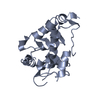
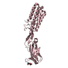
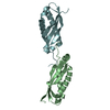
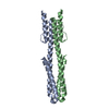
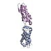
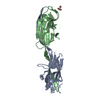
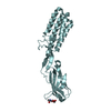
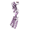
| Title | Crystal structure of Izumo1, the mammalian sperm ligand for egg Juno | |||||||||||||||||||||
 Components Components | Izumo sperm-egg fusion protein 1 | |||||||||||||||||||||
 Keywords Keywords | CELL ADHESION / FERTILIZATION / EGG RECEPTOR / GAMETE ADHESION / SPERM-EGG MEMBRANE FUSION | |||||||||||||||||||||
| Function / homology |  Function and homology information Function and homology informationAcrosome Reaction and Sperm:Oocyte Membrane Binding / protein complex involved in cell-cell adhesion / syncytium formation by cell-cell fusion / sperm-egg recognition / protein binding involved in heterotypic cell-cell adhesion / fusion of sperm to egg plasma membrane involved in single fertilization / acrosomal membrane / binding of sperm to zona pellucida / acrosomal vesicle / cell adhesion ...Acrosome Reaction and Sperm:Oocyte Membrane Binding / protein complex involved in cell-cell adhesion / syncytium formation by cell-cell fusion / sperm-egg recognition / protein binding involved in heterotypic cell-cell adhesion / fusion of sperm to egg plasma membrane involved in single fertilization / acrosomal membrane / binding of sperm to zona pellucida / acrosomal vesicle / cell adhesion / receptor ligand activity / signaling receptor binding / endoplasmic reticulum membrane / protein homodimerization activity / membrane / identical protein binding / plasma membrane Similarity search - Function | |||||||||||||||||||||
| Biological species |  | |||||||||||||||||||||
| Method |  X-RAY DIFFRACTION / SYNCHROTRON / MOLECULAR REPLACEMENT / Resolution: 2.5 Å X-RAY DIFFRACTION / SYNCHROTRON / MOLECULAR REPLACEMENT / Resolution: 2.5 Å | |||||||||||||||||||||
 Authors Authors | Nishimura, K. / Han, L. / De Sanctis, D. / Jovine, L. | |||||||||||||||||||||
| Funding support |  Sweden, 6items Sweden, 6items
| |||||||||||||||||||||
 Citation Citation | Journal: Curr.Biol. / Year: 2016 Title: The structure of sperm Izumo1 reveals unexpected similarities with Plasmodium invasion proteins. Authors: Nishimura, K. / Han, L. / Bianchi, E. / Wright, G.J. / de Sanctis, D. / Jovine, L. #1: Journal: Curr. Biol. / Year: 2016Title: Divergent evolution of vitamin B9 binding underlies Juno-mediated adhesion of mammalian gametes. Authors: Han, L. / Nishimura, K. / Sadat Al Hosseini, H. / Bianchi, E. / Wright, G.J. / Jovine, L. #2: Journal: Nature / Year: 2014 Title: Juno is the egg Izumo receptor and is essential for mammalian fertilization. Authors: Bianchi, E. / Doe, B. / Goulding, D. / Wright, G.J. #3: Journal: Nature / Year: 2005 Title: The immunoglobulin superfamily protein Izumo is required for sperm to fuse with eggs. Authors: Inoue, N. / Ikawa, M. / Isotani, A. / Okabe, M. | |||||||||||||||||||||
| History |
|
- Structure visualization
| Structure viewer | Molecule: MolmilJmol/JSmol |
|---|
- Downloads & links
-Download
| PDBx/mmCIF format | 5b5k.cif.gz | 151.7 KB | Display | PDBx/mmCIF format |
|---|---|---|---|---|
| PDB format | pdb5b5k.ent.gz | 121.9 KB | Display | PDB format |
| PDBx/mmJSON format | 5b5k.json.gz | Tree view | PDBx/mmJSON format | |
| Others |  Other downloads Other downloads |
-Validation report
| Arichive directory | https://data.pdbj.org/pub/pdb/validation_reports/b5/5b5kftp://data.pdbj.org/pub/pdb/validation_reports/b5/5b5k | HTTPS FTP |
|---|
-Related structure data
| Related structure data |  5jk9S S: Starting model for refinement |
|---|---|
| Similar structure data |


-Links
 PDBj
PDBj- Assembly
| Deposited unit | 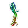
| ||||||||
|---|---|---|---|---|---|---|---|---|---|
| 1 |
| ||||||||
| Unit cell |
|
-Components
| #1: Protein | Mass: 27332.318 Da / Num. of mol.: 1 / Fragment: UNP RESIDUES 22-257 Source method: isolated from a genetically manipulated source Source: (gene. exp.)  Homo sapiens (human) / References: UniProt: Q9D9J7 Homo sapiens (human) / References: UniProt: Q9D9J7 |
|---|---|
| #2: Sugar | ChemComp-NAG /   Type: D-saccharide, beta linking / Mass: 221.208 Da / Num. of mol.: 1 Type: D-saccharide, beta linking / Mass: 221.208 Da / Num. of mol.: 1Source method: isolated from a genetically manipulated source Formula: C8H15NO6 |
| #3: Chemical | ChemComp-EPE /   Mass: 238.305 Da / Num. of mol.: 1 Mass: 238.305 Da / Num. of mol.: 1Source method: isolated from a genetically manipulated source Formula: C8H18N2O4S / Comment: pH buffer*YM |
| #4: Water | ChemComp-HOH /  Mass: 18.015 Da / Num. of mol.: 28 / Source method: isolated from a natural source / Formula: H2O Mass: 18.015 Da / Num. of mol.: 28 / Source method: isolated from a natural source / Formula: H2O |
| Has protein modification | Y |
-Experimental details
-Experiment
| Experiment | Method: X-RAY DIFFRACTION / Number of used crystals: 1 |
|---|
- Sample preparation
| Crystal | Density Matthews: 3.47 Å3/Da / Density % sol: 64.6 % / Description: Square plate |
|---|---|
| Crystal grow | Temperature: 293 K / Method: vapor diffusion, sitting drop / pH: 6.6 / Details: 0.2 M Ammonium formate pH 6.6, 20% PEG 3350 |
-Data collection
| Diffraction | Mean temperature: 100 K |
|---|---|
| Diffraction source | Source: SYNCHROTRON / Site: ESRF  / Beamline: ID29 / Wavelength: 0.984 Å / Beamline: ID29 / Wavelength: 0.984 Å |
| Detector | Type: DECTRIS PILATUS 6M-F / Detector: PIXEL / Date: May 14, 2015 |
| Radiation | Protocol: SINGLE WAVELENGTH / Monochromatic (M) / Laue (L): M / Scattering type: x-ray |
| Radiation wavelength | Wavelength: 0.984 Å / Relative weight: 1 |
| Reflection | Resolution: 2.5→43.84 Å / Num. obs: 13389 / % possible obs: 100 % / Redundancy: 12.6 % / Biso Wilson estimate: 59.2 Å2 / CC1/2: 0.997 / Rmerge(I) obs: 0.1948 / Net I/σ(I): 12.13 |
| Reflection shell | Resolution: 2.5→2.589 Å / Redundancy: 10.1 % / Rmerge(I) obs: 0.2296 / Mean I/σ(I) obs: 1.13 / % possible all: 100 |
- Processing
| Software |
| |||||||||||||||||||||||||||||||||||||||||||||||||||||||||||||||||||||||||||
|---|---|---|---|---|---|---|---|---|---|---|---|---|---|---|---|---|---|---|---|---|---|---|---|---|---|---|---|---|---|---|---|---|---|---|---|---|---|---|---|---|---|---|---|---|---|---|---|---|---|---|---|---|---|---|---|---|---|---|---|---|---|---|---|---|---|---|---|---|---|---|---|---|---|---|---|---|
| Refinement | Method to determine structure: MOLECULAR REPLACEMENT Starting model: 5JK9 Resolution: 2.5→43.84 Å / SU ML: 0.46 / Cross valid method: FREE R-VALUE / σ(F): 1.35 / Phase error: 31.93
| |||||||||||||||||||||||||||||||||||||||||||||||||||||||||||||||||||||||||||
| Solvent computation | Shrinkage radii: 0.9 Å / VDW probe radii: 1.11 Å | |||||||||||||||||||||||||||||||||||||||||||||||||||||||||||||||||||||||||||
| Refinement step | Cycle: LAST / Resolution: 2.5→43.84 Å
| |||||||||||||||||||||||||||||||||||||||||||||||||||||||||||||||||||||||||||
| Refine LS restraints |
| |||||||||||||||||||||||||||||||||||||||||||||||||||||||||||||||||||||||||||
| LS refinement shell |
| |||||||||||||||||||||||||||||||||||||||||||||||||||||||||||||||||||||||||||
| Refinement TLS params. | Method: refined / Refine-ID: X-RAY DIFFRACTION
| |||||||||||||||||||||||||||||||||||||||||||||||||||||||||||||||||||||||||||
| Refinement TLS group |
|
