 Movie
Movie Controller

[English] 日本語
 Yorodumi
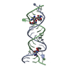
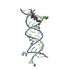
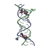
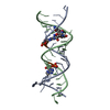
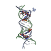
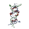
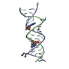
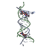
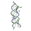
- PDB-3td1: Crystal structure of the bacterial A1408G-mutant and the protozoa... -
+ Open data

- Basic information
| Entry | Database: PDB / ID: 3td1 | ||||||||||||||||||
|---|---|---|---|---|---|---|---|---|---|---|---|---|---|---|---|---|---|---|---|
| Title | Crystal structure of the bacterial A1408G-mutant and the protozoa cytoplasmic ribosomal decoding site in complex with geneticin | ||||||||||||||||||
 Components Components | RNA (5'-R(* Keywords KeywordsRNA/ANTIBIOTIC / decoding / ribosome / RNA-ANTIBIOTIC complex | Function / homology | GENETICIN / RNA / RNA (> 10) |  Function and homology information Function and homology informationMethod |  X-RAY DIFFRACTION / SYNCHROTRON / MOLECULAR REPLACEMENT / Resolution: 2.1 Å X-RAY DIFFRACTION / SYNCHROTRON / MOLECULAR REPLACEMENT / Resolution: 2.1 Å  Authors AuthorsKondo, J. |  CitationJournal: Angew.Chem.Int.Ed.Engl. / Year: 2012 CitationJournal: Angew.Chem.Int.Ed.Engl. / Year: 2012Title: A structural basis for the antibiotic resistance conferred by an A1408G mutation in 16S rRNA and for the antiprotozoal activity of aminoglycosides Authors: Kondo, J. History |
|
- Structure visualization
| Structure viewer | Molecule: MolmilJmol/JSmol |
|---|
- Downloads & links
-Download
| PDBx/mmCIF format | 3td1.cif.gz | 42 KB | Display | PDBx/mmCIF format |
|---|---|---|---|---|
| PDB format | pdb3td1.ent.gz | 29 KB | Display | PDB format |
| PDBx/mmJSON format | 3td1.json.gz | Tree view | PDBx/mmJSON format | |
| Others |  Other downloads Other downloads |
-Validation report
| Summary document | 3td1_validation.pdf.gz | 1.1 MB | Display | wwPDB validaton report |
|---|---|---|---|---|
| Full document | 3td1_full_validation.pdf.gz | 1.1 MB | Display | |
| Data in XML | 3td1_validation.xml.gz | 6.6 KB | Display | |
| Data in CIF | 3td1_validation.cif.gz | 9.3 KB | Display | |
| Arichive directory | https://data.pdbj.org/pub/pdb/validation_reports/td/3td1ftp://data.pdbj.org/pub/pdb/validation_reports/td/3td1 | HTTPS FTP |
-Related structure data
| Related structure data | 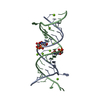 3td0C 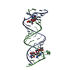 1mwlS S: Starting model for refinement C: citing same article ( |
|---|---|
| Similar structure data |

-Links
 PDBj
PDBj


- Assembly
| Deposited unit | 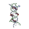
| |||||||||
|---|---|---|---|---|---|---|---|---|---|---|
| 1 |
| |||||||||
| Unit cell |
| |||||||||
| Components on special symmetry positions |
|
-Components
| #1: RNA chain | Mass: 7450.304 Da / Num. of mol.: 2 / Source method: obtained synthetically / Details: Synthetic RNA #2: Chemical |   Mass: 496.552 Da / Num. of mol.: 2 / Source method: obtained synthetically / Formula: C20H40N4O10 / Comment: antibiotic*YM Mass: 496.552 Da / Num. of mol.: 2 / Source method: obtained synthetically / Formula: C20H40N4O10 / Comment: antibiotic*YM#3: Water | ChemComp-HOH / |  Mass: 18.015 Da / Num. of mol.: 189 / Source method: isolated from a natural source / Formula: H2O Mass: 18.015 Da / Num. of mol.: 189 / Source method: isolated from a natural source / Formula: H2O |
|---|
-Experimental details
-Experiment
| Experiment | Method: X-RAY DIFFRACTION / Number of used crystals: 1 |
|---|
- Sample preparation
| Crystal | Density Matthews: 2.29 Å3/Da / Density % sol: 46.32 % |
|---|---|
| Crystal grow | Temperature: 293 K / Method: vapor diffusion, hanging drop / pH: 7 Details: Sodium Cacodylate, Spermine tetrahydrochloride, Ammonium chloride, 2-methyl-2,4-pentanediol , pH 7.0, VAPOR DIFFUSION, HANGING DROP, temperature 293K |
-Data collection
| Diffraction | Mean temperature: 100 K |
|---|---|
| Diffraction source | Source: SYNCHROTRON / Site: Photon Factory  / Beamline: BL-17A / Wavelength: 1 Å / Beamline: BL-17A / Wavelength: 1 Å |
| Detector | Type: ADSC QUANTUM 315r / Detector: CCD / Date: Feb 26, 2011 |
| Radiation | Protocol: SINGLE WAVELENGTH / Monochromatic (M) / Laue (L): M / Scattering type: x-ray |
| Radiation wavelength | Wavelength: 1 Å / Relative weight: 1 |
| Reflection | Resolution: 2.1→31.9 Å / Num. obs: 8461 / % possible obs: 99.9 % / Redundancy: 6.9 % / Rmerge(I) obs: 0.065 |
| Reflection shell | Resolution: 2.1→2.2 Å / Redundancy: 7 % / Rmerge(I) obs: 0.359 / % possible all: 100 |
- Processing
| Software |
| ||||||||||||||||
|---|---|---|---|---|---|---|---|---|---|---|---|---|---|---|---|---|---|
| Refinement | Method to determine structure: MOLECULAR REPLACEMENT Starting model: PDB entry 1MWL Resolution: 2.1→31.9 Å
| ||||||||||||||||
| Refinement step | Cycle: LAST / Resolution: 2.1→31.9 Å
| ||||||||||||||||
| Refine LS restraints |
|
