 Movie
Movie Controller

[English] 日本語
 Yorodumi
- PDB-5z1i: Crystal structure of the protozoal cytoplasmic ribosomal decoding... -
+ Open data

- Basic information
| Entry | Database: PDB / ID: 5z1i | ||||||||||||||||||||||||||||
|---|---|---|---|---|---|---|---|---|---|---|---|---|---|---|---|---|---|---|---|---|---|---|---|---|---|---|---|---|---|
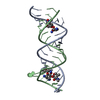
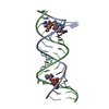
| Title | Crystal structure of the protozoal cytoplasmic ribosomal decoding site in complex with 6'-fluoro sisomicin | ||||||||||||||||||||||||||||
 Components Components | RNA (5'-R(P* Keywords KeywordsRNA / rRNA / aminoglycoside | Function / homology | Chem-FSJ / RNA / RNA (> 10) |  Function and homology information Function and homology informationBiological species | synthetic construct (others) | Method |  X-RAY DIFFRACTION / SYNCHROTRON / Resolution: 1.903 Å X-RAY DIFFRACTION / SYNCHROTRON / Resolution: 1.903 Å  Authors AuthorsKanazawa, H. / Hanessian, S. / Kondo, J. | Funding support | |  Japan, 1items Japan, 1items
 CitationJournal: ChemMedChem / Year: 2018 CitationJournal: ChemMedChem / Year: 2018Title: Structure-Based Design of a Eukaryote-Selective Antiprotozoal Fluorinated Aminoglycoside. Authors: Kanazawa, H. / Saavedra, O.M. / Maianti, J.P. / Young, S.A. / Izquierdo, L. / Smith, T.K. / Hanessian, S. / Kondo, J. History |
|
- Structure visualization
| Structure viewer | Molecule: MolmilJmol/JSmol |
|---|
- Downloads & links
-Download
| PDBx/mmCIF format | 5z1i.cif.gz | 26.9 KB | Display | PDBx/mmCIF format |
|---|---|---|---|---|
| PDB format | pdb5z1i.ent.gz | 15.8 KB | Display | PDB format |
| PDBx/mmJSON format | 5z1i.json.gz | Tree view | PDBx/mmJSON format | |
| Others |  Other downloads Other downloads |
-Validation report
| Arichive directory | https://data.pdbj.org/pub/pdb/validation_reports/z1/5z1iftp://data.pdbj.org/pub/pdb/validation_reports/z1/5z1i | HTTPS FTP |
|---|
-Related structure data

-Links
 PDBj
PDBj


- Assembly
| Deposited unit | 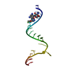
| ||||||||
|---|---|---|---|---|---|---|---|---|---|
| 1 | 
| ||||||||
| Unit cell |
| ||||||||
| Components on special symmetry positions |
|
-Components
| #1: RNA chain | Mass: 6774.093 Da / Num. of mol.: 1 / Source method: obtained synthetically / Source: (synth.) synthetic construct (others) |
|---|---|
| #2: Chemical | ChemComp-FSJ / (  Mass: 450.502 Da / Num. of mol.: 1 Mass: 450.502 Da / Num. of mol.: 1Source method: isolated from a genetically manipulated source Formula: C19H35FN4O7 |
| #3: Water | ChemComp-HOH /  Mass: 18.015 Da / Num. of mol.: 73 / Source method: isolated from a natural source / Formula: H2O Mass: 18.015 Da / Num. of mol.: 73 / Source method: isolated from a natural source / Formula: H2O |
-Experimental details
-Experiment
| Experiment | Method: X-RAY DIFFRACTION / Number of used crystals: 1 |
|---|
- Sample preparation
| Crystal | Density Matthews: 2.27 Å3/Da / Density % sol: 45.76 % |
|---|---|
| Crystal grow | Temperature: 293 K / Method: vapor diffusion, hanging drop / pH: 7 Details: 2-Methyl-2,4-pentanediol, LiCl, spermine tetrahydrochloride, sodium cacodylate |
-Data collection
| Diffraction | Mean temperature: 100 K | ||||||||||||||||||||||||||||||||||||||||||||||||||||||||||||||||||||||||||||||||||||||||||||||||||||||||||||||||||||||||||||||||||||||||||||||||||||||||||||||||||||||||
|---|---|---|---|---|---|---|---|---|---|---|---|---|---|---|---|---|---|---|---|---|---|---|---|---|---|---|---|---|---|---|---|---|---|---|---|---|---|---|---|---|---|---|---|---|---|---|---|---|---|---|---|---|---|---|---|---|---|---|---|---|---|---|---|---|---|---|---|---|---|---|---|---|---|---|---|---|---|---|---|---|---|---|---|---|---|---|---|---|---|---|---|---|---|---|---|---|---|---|---|---|---|---|---|---|---|---|---|---|---|---|---|---|---|---|---|---|---|---|---|---|---|---|---|---|---|---|---|---|---|---|---|---|---|---|---|---|---|---|---|---|---|---|---|---|---|---|---|---|---|---|---|---|---|---|---|---|---|---|---|---|---|---|---|---|---|---|---|---|---|
| Diffraction source | Source: SYNCHROTRON / Site: Photon Factory / Beamline: BL-17A / Wavelength: 0.98 Å | ||||||||||||||||||||||||||||||||||||||||||||||||||||||||||||||||||||||||||||||||||||||||||||||||||||||||||||||||||||||||||||||||||||||||||||||||||||||||||||||||||||||||
| Detector | Type: ADSC QUANTUM 270 / Detector: CCD / Date: Mar 6, 2016 | ||||||||||||||||||||||||||||||||||||||||||||||||||||||||||||||||||||||||||||||||||||||||||||||||||||||||||||||||||||||||||||||||||||||||||||||||||||||||||||||||||||||||
| Radiation | Protocol: SINGLE WAVELENGTH / Monochromatic (M) / Laue (L): M / Scattering type: x-ray | ||||||||||||||||||||||||||||||||||||||||||||||||||||||||||||||||||||||||||||||||||||||||||||||||||||||||||||||||||||||||||||||||||||||||||||||||||||||||||||||||||||||||
| Radiation wavelength | Wavelength: 0.98 Å / Relative weight: 1 | ||||||||||||||||||||||||||||||||||||||||||||||||||||||||||||||||||||||||||||||||||||||||||||||||||||||||||||||||||||||||||||||||||||||||||||||||||||||||||||||||||||||||
| Reflection | Resolution: 1.9→42.862 Å / Num. obs: 5236 / % possible obs: 99.8 % / Redundancy: 11.955 % / Biso Wilson estimate: 20.85 Å2 / CC1/2: 0.999 / Rmerge(I) obs: 0.072 / Rrim(I) all: 0.075 / Χ2: 1.044 / Net I/σ(I): 23.85 | ||||||||||||||||||||||||||||||||||||||||||||||||||||||||||||||||||||||||||||||||||||||||||||||||||||||||||||||||||||||||||||||||||||||||||||||||||||||||||||||||||||||||
| Reflection shell | Diffraction-ID: 1
|
- Processing
| Software |
| ||||||||||||||||||||||||||||||||||||||||||||||||||||||||
|---|---|---|---|---|---|---|---|---|---|---|---|---|---|---|---|---|---|---|---|---|---|---|---|---|---|---|---|---|---|---|---|---|---|---|---|---|---|---|---|---|---|---|---|---|---|---|---|---|---|---|---|---|---|---|---|---|---|
| Refinement | Resolution: 1.903→42.862 Å / SU ML: 0.15 / Cross valid method: THROUGHOUT / σ(F): 1.35 / Phase error: 19.82
| ||||||||||||||||||||||||||||||||||||||||||||||||||||||||
| Solvent computation | Shrinkage radii: 0.9 Å / VDW probe radii: 1.11 Å | ||||||||||||||||||||||||||||||||||||||||||||||||||||||||
| Displacement parameters | Biso max: 198.25 Å2 / Biso mean: 25.48 Å2 / Biso min: 12.5 Å2 | ||||||||||||||||||||||||||||||||||||||||||||||||||||||||
| Refinement step | Cycle: final / Resolution: 1.903→42.862 Å
| ||||||||||||||||||||||||||||||||||||||||||||||||||||||||
| Refine LS restraints |
| ||||||||||||||||||||||||||||||||||||||||||||||||||||||||
| LS refinement shell | Refine-ID: X-RAY DIFFRACTION / Rfactor Rfree error: 0 / Total num. of bins used: 7
|
