 Movie
Movie Controller

[English] 日本語
 Yorodumi
- PDB-3t7r: Crystal structure of apo BVU_3255, a methyltransferase from Bacte... -
+ Open data

- Basic information
| Entry | Database: PDB / ID: 3t7r | ||||||
|---|---|---|---|---|---|---|---|
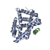
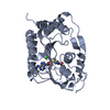
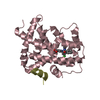
| Title | Crystal structure of apo BVU_3255, a methyltransferase from Bacteroides vulgatus ATCC 8482 | ||||||
 Components Components | Putative methyltransferase | ||||||
 Keywords Keywords | TRANSFERASE / Small molecule methyltransferase / BVU_3255 / ROSSMANN FOLD / METHYLTRASNFERASE / SAM / Methylation | ||||||
| Function / homology |  Function and homology information Function and homology information | ||||||
| Biological species |  Bacteroides vulgatus (bacteria) Bacteroides vulgatus (bacteria) | ||||||
| Method |  X-RAY DIFFRACTION / MOLECULAR REPLACEMENT / Resolution: 2.9 Å X-RAY DIFFRACTION / MOLECULAR REPLACEMENT / Resolution: 2.9 Å | ||||||
 Authors Authors | Kumar, V. / Sivaraman, J. | ||||||
 Citation Citation | Journal: J.Struct.Biol. / Year: 2011 Title: Structural characterization of BVU_3255, a methyltransferase from human intestine antibiotic resistant pathogen Bacteroides vulgatus Authors: Kumar, V. / Sivaraman, J. | ||||||
| History |
|
- Structure visualization
| Structure viewer | Molecule: MolmilJmol/JSmol |
|---|
- Downloads & links
-Download
| PDBx/mmCIF format | 3t7r.cif.gz | 112 KB | Display | PDBx/mmCIF format |
|---|---|---|---|---|
| PDB format | pdb3t7r.ent.gz | 86.2 KB | Display | PDB format |
| PDBx/mmJSON format | 3t7r.json.gz | Tree view | PDBx/mmJSON format | |
| Others |  Other downloads Other downloads |
-Validation report
| Summary document | 3t7r_validation.pdf.gz | 461.4 KB | Display | wwPDB validaton report |
|---|---|---|---|---|
| Full document | 3t7r_full_validation.pdf.gz | 471.8 KB | Display | |
| Data in XML | 3t7r_validation.xml.gz | 22.2 KB | Display | |
| Data in CIF | 3t7r_validation.cif.gz | 29.7 KB | Display | |
| Arichive directory | https://data.pdbj.org/pub/pdb/validation_reports/t7/3t7rftp://data.pdbj.org/pub/pdb/validation_reports/t7/3t7r | HTTPS FTP |
-Related structure data
| Related structure data |  3t7sC  3t7tC  3e7pS S: Starting model for refinement C: citing same article ( |
|---|---|
| Similar structure data |


-Links
 PDBj
PDBj- Assembly
| Deposited unit | 
| ||||||||
|---|---|---|---|---|---|---|---|---|---|
| 1 | 
| ||||||||
| 2 | 
| ||||||||
| Unit cell |
|
-Components
| #1: Protein | Mass: 30664.715 Da / Num. of mol.: 2 Source method: isolated from a genetically manipulated source Source: (gene. exp.) Bacteroides vulgatus (bacteria) / Strain: ATCC 8482 / Gene: BVU_3255 / Plasmid: pGS21a / Production host: #2: Chemical |   Mass: 192.254 Da / Num. of mol.: 2 / Source method: obtained synthetically / Formula: C12H16O2 Mass: 192.254 Da / Num. of mol.: 2 / Source method: obtained synthetically / Formula: C12H16O2#3: Water | ChemComp-HOH / |  Mass: 18.015 Da / Num. of mol.: 101 / Source method: isolated from a natural source / Formula: H2O Mass: 18.015 Da / Num. of mol.: 101 / Source method: isolated from a natural source / Formula: H2ONonpolymer details | THE LIGAND 6PP HAS BEEN BUILT BASED ON THE DENSITY OF THIS STRUCTURE. THE ORIGINAL COMPOUND MIGHT ...THE LIGAND 6PP HAS BEEN BUILT BASED ON THE DENSITY OF THIS STRUCTURE. THE ORIGINAL COMPOUND MIGHT BE 2-POLYPRENYL | |
|---|
-Experimental details
-Experiment
| Experiment | Method: X-RAY DIFFRACTION / Number of used crystals: 1 |
|---|
- Sample preparation
| Crystal | Density Matthews: 2.17 Å3/Da / Density % sol: 43.33 % |
|---|---|
| Crystal grow | Temperature: 298 K / Method: vapor diffusion, hanging drop Details: 170mM Mg(CH3COO)2, 15% Polyethylene Glycol 3350, VAPOR DIFFUSION, HANGING DROP, temperature 298K |
-Data collection
| Diffraction | Mean temperature: 100 K |
|---|---|
| Diffraction source | Source: ROTATING ANODE / Type: BRUKER AXS MICROSTAR / Wavelength: 1.5418 Å |
| Detector | Type: Bruker Platinum 135 / Detector: CCD / Date: Mar 11, 2011 |
| Radiation | Monochromator: Graphite / Protocol: SINGLE WAVELENGTH / Monochromatic (M) / Laue (L): M / Scattering type: x-ray |
| Radiation wavelength | Wavelength: 1.5418 Å / Relative weight: 1 |
| Reflection | Resolution: 2.9→50 Å / Num. obs: 11578 / % possible obs: 92.8 % / Observed criterion σ(F): 1 / Observed criterion σ(I): 1 / Rsym value: 0.105 / Net I/σ(I): 23.3 |
| Reflection shell | Resolution: 2.9→3 Å / Mean I/σ(I) obs: 4.8 / Rsym value: 0.184 / % possible all: 74.5 |
- Processing
| Software |
| ||||||||||||||||||||||||||||
|---|---|---|---|---|---|---|---|---|---|---|---|---|---|---|---|---|---|---|---|---|---|---|---|---|---|---|---|---|---|
| Refinement | Method to determine structure: MOLECULAR REPLACEMENT Starting model: PDB ENTRY 3E7P Resolution: 2.9→30 Å / Occupancy max: 1 / Occupancy min: 1 / Isotropic thermal model: Isotropic / Cross valid method: THROUGHOUT / σ(F): 2 / Stereochemistry target values: Engh & Huber
| ||||||||||||||||||||||||||||
| Solvent computation | Bsol: 8.2466 Å2 | ||||||||||||||||||||||||||||
| Displacement parameters | Biso max: 67.07 Å2 / Biso mean: 40.849 Å2 / Biso min: 1 Å2
| ||||||||||||||||||||||||||||
| Refinement step | Cycle: LAST / Resolution: 2.9→30 Å
| ||||||||||||||||||||||||||||
| Refine LS restraints |
| ||||||||||||||||||||||||||||
| Xplor file |
|
