 Movie
Movie Controller


+ Open data

- Basic information
| Entry | Database: PDB / ID: 3t5d | ||||||
|---|---|---|---|---|---|---|---|
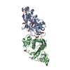
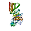
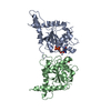
| Title | Crystal structure of Septin 7 in complex with GDP | ||||||
 Components Components | Septin-7 | ||||||
 Keywords Keywords | SIGNALING PROTEIN / GTP-binding protein / cytoskeleton | ||||||
| Function / homology |  Function and homology information Function and homology informationregulation of embryonic cell shape / positive regulation of non-motile cilium assembly / septin complex / sperm annulus / cytoskeleton-dependent cytokinesis / septin ring / non-motile cilium / cell division site / cleavage furrow / cilium assembly ...regulation of embryonic cell shape / positive regulation of non-motile cilium assembly / septin complex / sperm annulus / cytoskeleton-dependent cytokinesis / septin ring / non-motile cilium / cell division site / cleavage furrow / cilium assembly / axoneme / stress fiber / MAPK6/MAPK4 signaling / spindle / kinetochore / intracellular protein localization / microtubule cytoskeleton / midbody / spermatogenesis / molecular adaptor activity / cell differentiation / cadherin binding / GTPase activity / GTP binding / structural molecule activity / extracellular exosome / identical protein binding / nucleus / cytosol Similarity search - Function | ||||||
| Biological species |  Homo sapiens (human) Homo sapiens (human) | ||||||
| Method |  X-RAY DIFFRACTION / SYNCHROTRON / MOLECULAR REPLACEMENT / Resolution: 3.3 Å X-RAY DIFFRACTION / SYNCHROTRON / MOLECULAR REPLACEMENT / Resolution: 3.3 Å | ||||||
 Authors Authors | Zent, E. / Wittinghofer, A. | ||||||
 Citation Citation | Journal: Biol.Chem. / Year: 2011 Title: Structural and biochemical properties of Sept7, a unique septin required for filament formation. Authors: Zent, E. / Vetter, I. / Wittinghofer, A. | ||||||
| History |
|
- Structure visualization
| Structure viewer | Molecule: MolmilJmol/JSmol |
|---|
- Downloads & links
-Download
| PDBx/mmCIF format | 3t5d.cif.gz | 105.7 KB | Display | PDBx/mmCIF format |
|---|---|---|---|---|
| PDB format | pdb3t5d.ent.gz | 81.4 KB | Display | PDB format |
| PDBx/mmJSON format | 3t5d.json.gz | Tree view | PDBx/mmJSON format | |
| Others |  Other downloads Other downloads |
-Validation report
| Arichive directory | https://data.pdbj.org/pub/pdb/validation_reports/t5/3t5dftp://data.pdbj.org/pub/pdb/validation_reports/t5/3t5d | HTTPS FTP |
|---|
-Related structure data
| Similar structure data |
|---|


-Links
 PDBj
PDBj- Assembly
| Deposited unit | 
| ||||||||||||||||||
|---|---|---|---|---|---|---|---|---|---|---|---|---|---|---|---|---|---|---|---|
| 1 |
| ||||||||||||||||||
| Unit cell |
| ||||||||||||||||||
| Noncrystallographic symmetry (NCS) | NCS domain:
NCS domain segments: Component-ID: 1 / Ens-ID: 1 / Beg auth comp-ID: PHE / Beg label comp-ID: PHE / End auth comp-ID: LEU / End label comp-ID: LEU / Refine code: 2 / Auth seq-ID: 30 - 295 / Label seq-ID: 7 - 272
|
-Components
| #1: Protein | Mass: 31349.900 Da / Num. of mol.: 2 Source method: isolated from a genetically manipulated source Source: (gene. exp.) Homo sapiens (human) / Gene: SEPT7, CDC10 / Production host:  #2: Chemical |   Type: RNA linking / Mass: 443.201 Da / Num. of mol.: 2 / Source method: obtained synthetically / Formula: C10H15N5O11P2 / Comment: GDP, energy-carrying molecule*YM Type: RNA linking / Mass: 443.201 Da / Num. of mol.: 2 / Source method: obtained synthetically / Formula: C10H15N5O11P2 / Comment: GDP, energy-carrying molecule*YM#3: Water | ChemComp-HOH / |  Mass: 18.015 Da / Num. of mol.: 2 / Source method: isolated from a natural source / Formula: H2O Mass: 18.015 Da / Num. of mol.: 2 / Source method: isolated from a natural source / Formula: H2O |
|---|
-Experimental details
-Experiment
| Experiment | Method: X-RAY DIFFRACTION / Number of used crystals: 1 |
|---|
- Sample preparation
| Crystal | Density Matthews: 3.29 Å3/Da / Density % sol: 62.6 % |
|---|---|
| Crystal grow | Temperature: 293 K / Method: vapor diffusion, hanging drop / pH: 9.5 Details: 0.2M sodium chlorid, 1.1M ammonium sulfate, 0.1M CHES, pH 9.5, VAPOR DIFFUSION, HANGING DROP, temperature 293K |
-Data collection
| Diffraction | Mean temperature: 100 K |
|---|---|
| Diffraction source | Source: SYNCHROTRON / Site: SLS  / Beamline: X10SA / Wavelength: 0.99986 Å / Beamline: X10SA / Wavelength: 0.99986 Å |
| Detector | Type: DECTRIS PILATUS 6M / Detector: PIXEL / Date: Aug 14, 2009 |
| Radiation | Protocol: SINGLE WAVELENGTH / Monochromatic (M) / Laue (L): M / Scattering type: x-ray |
| Radiation wavelength | Wavelength: 0.99986 Å / Relative weight: 1 |
| Reflection | Resolution: 3.3→71.25 Å / Num. all: 12190 / Num. obs: 12079 / % possible obs: 99.1 % / Redundancy: 8.9 % |
| Reflection shell | Resolution: 3.3→3.4 Å / Redundancy: 9.1 % / Rmerge(I) obs: 0.392 / Mean I/σ(I) obs: 3.85 / Num. unique all: 1007 / Rsym value: 0.596 / % possible all: 98.9 |
- Processing
| Software |
| ||||||||||||||||||||||||||||||||||||||||||||||||||||||||||||||||||||||||||||||||||||||||||
|---|---|---|---|---|---|---|---|---|---|---|---|---|---|---|---|---|---|---|---|---|---|---|---|---|---|---|---|---|---|---|---|---|---|---|---|---|---|---|---|---|---|---|---|---|---|---|---|---|---|---|---|---|---|---|---|---|---|---|---|---|---|---|---|---|---|---|---|---|---|---|---|---|---|---|---|---|---|---|---|---|---|---|---|---|---|---|---|---|---|---|---|
| Refinement | Method to determine structure: MOLECULAR REPLACEMENT / Resolution: 3.3→71.25 Å / Cor.coef. Fo:Fc: 0.89 / Cor.coef. Fo:Fc free: 0.86 / SU B: 86.942 / SU ML: 0.653 / Cross valid method: THROUGHOUT / ESU R Free: 0.619 / Stereochemistry target values: MAXIMUM LIKELIHOOD / Details: HYDROGENS HAVE BEEN ADDED IN THE RIDING POSITIONS
| ||||||||||||||||||||||||||||||||||||||||||||||||||||||||||||||||||||||||||||||||||||||||||
| Solvent computation | Ion probe radii: 0.8 Å / Shrinkage radii: 0.8 Å / VDW probe radii: 1.2 Å / Solvent model: MASK | ||||||||||||||||||||||||||||||||||||||||||||||||||||||||||||||||||||||||||||||||||||||||||
| Displacement parameters | Biso mean: 68.02 Å2
| ||||||||||||||||||||||||||||||||||||||||||||||||||||||||||||||||||||||||||||||||||||||||||
| Refinement step | Cycle: LAST / Resolution: 3.3→71.25 Å
| ||||||||||||||||||||||||||||||||||||||||||||||||||||||||||||||||||||||||||||||||||||||||||
| Refine LS restraints |
| ||||||||||||||||||||||||||||||||||||||||||||||||||||||||||||||||||||||||||||||||||||||||||
| Refine LS restraints NCS | Dom-ID: 1 / Ens-ID: 1 / Refine-ID: X-RAY DIFFRACTION
| ||||||||||||||||||||||||||||||||||||||||||||||||||||||||||||||||||||||||||||||||||||||||||
| LS refinement shell | Resolution: 3.3→3.386 Å / Total num. of bins used: 20
|
