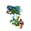
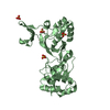
ジャーナル: Nat Struct Mol Biol / 年: 2011 タイトル: Structure of green-type Rubisco activase from tobacco. 著者: Mathias Stotz / Oliver Mueller-Cajar / Susanne Ciniawsky / Petra Wendler / F Ulrich Hartl / Andreas Bracher / Manajit Hayer-Hartl / 要旨: Rubisco, the enzyme that catalyzes the fixation of atmospheric CO(2) in photosynthesis, is subject to inactivation by inhibitory sugar phosphates. Here we report the 2.95-Å crystal structure of ...Rubisco, the enzyme that catalyzes the fixation of atmospheric CO(2) in photosynthesis, is subject to inactivation by inhibitory sugar phosphates. Here we report the 2.95-Å crystal structure of Nicotiana tabacum Rubisco activase (Rca), the enzyme that facilitates the removal of these inhibitors. Rca from tobacco has a classical AAA(+)-protein domain architecture. Although Rca populates a range of oligomeric states when in solution, it forms a helical arrangement with six subunits per turn when in the crystal. However, negative-stain electron microscopy of the active mutant R294V suggests that Rca functions as a hexamer. The residues determining species specificity for Rubisco are located in a helical insertion of the C-terminal domain and probably function in conjunction with the N-domain in Rubisco recognition. Loop segments exposed toward the central pore of the hexamer are required for the ATP-dependent remodeling of Rubisco, resulting in the release of inhibitory sugar.
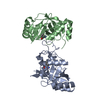
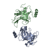
THE ANALYSIS OF THE CBBX PROTEIN IN SOLUTION AND EM STUDIES SUGGEST THAT THE BIOLOGICALLY ACTIVE OLIGOMER IS A HEXAMER, BUT IT CANNOT BE GENERATED BY THE APPLICATION OF SYMMETRY OPERATORS TO THE CHAINS IN THE COORDINATE FILE.
-
要素
#1: タンパク質
Ribulosebisphosphatecarboxylase/oxygenaseactivase1, chloroplastic / RA 1 / RuBisCO activase 1
プロトコル: SINGLE WAVELENGTH / 単色(M)・ラウエ(L): M / 散乱光タイプ: x-ray
放射波長
波長: 1.0332 Å / 相対比: 1
Reflection
冗長度: 5.5 % / Av σ(I) over netI: 9.7 / 数: 29140 / Rmerge(I) obs: 0.053 / Rsym value: 0.053 / D res high: 3.312 Å / D res low: 47.946 Å / Num. obs: 5298 / % possible obs: 99.7
 ムービー
ムービー コントローラー


 データを開く
 基本情報
基本情報 要素
要素 キーワード
キーワード 機能・相同性情報
 X線回折 /
  データ登録者
 引用
引用
 構造の表示
構造の表示 ダウンロードとリンク
 その他のダウンロード


 PDBj
PDBj 集合体
集合体

 試料調製
試料調製 / ビームライン: ID14-4 / 波長: 1.0332 Å
 解析
解析